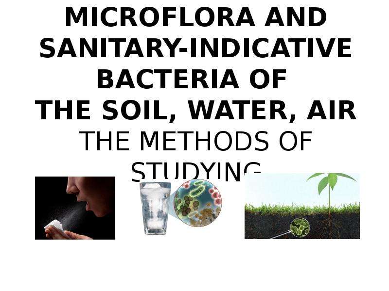
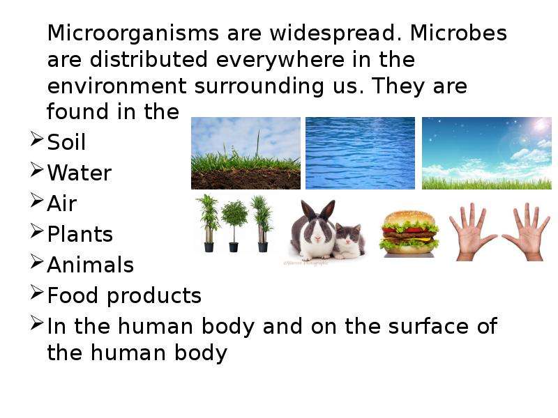
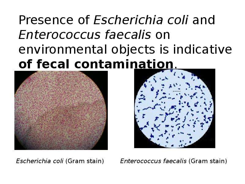
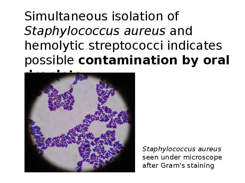
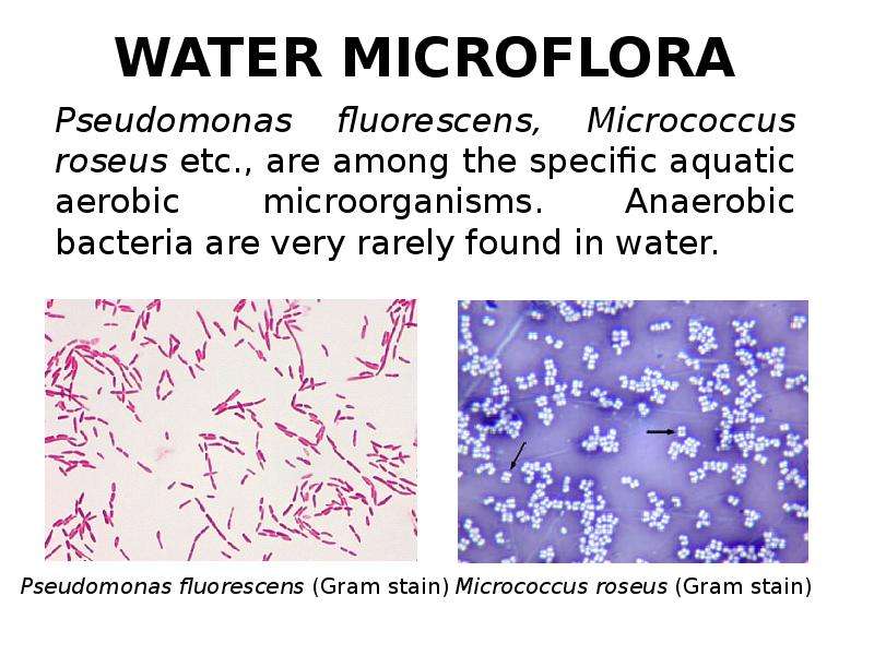
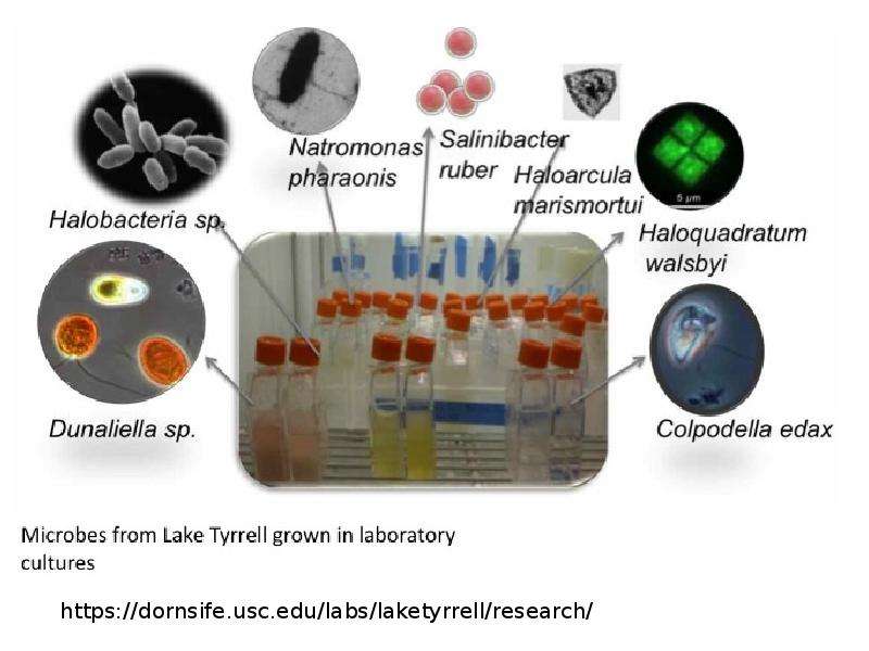
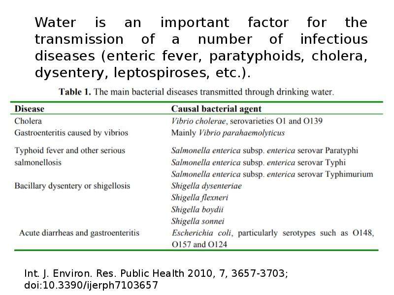
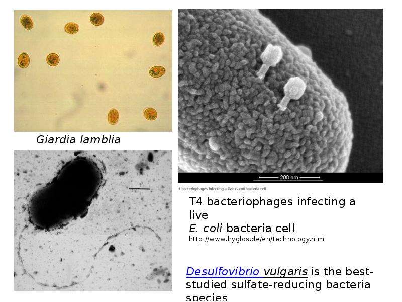
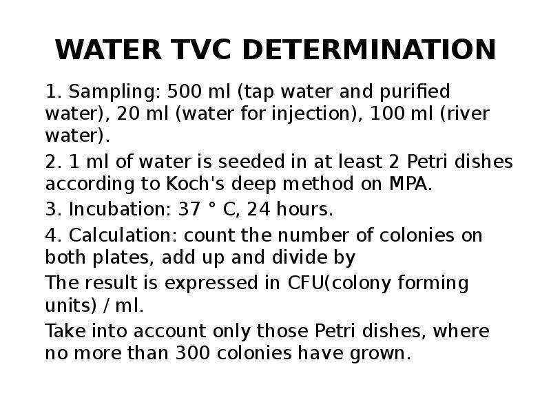
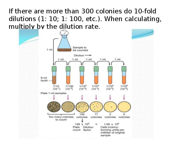
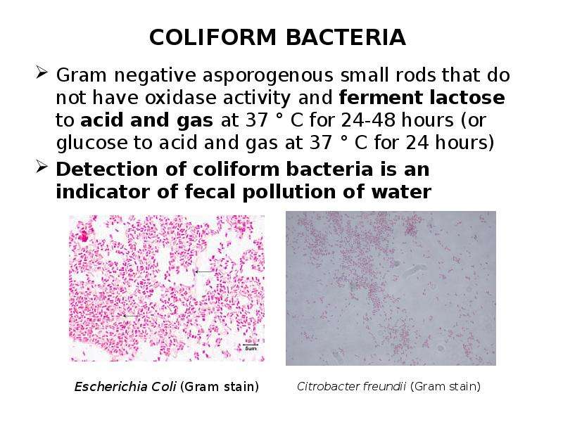
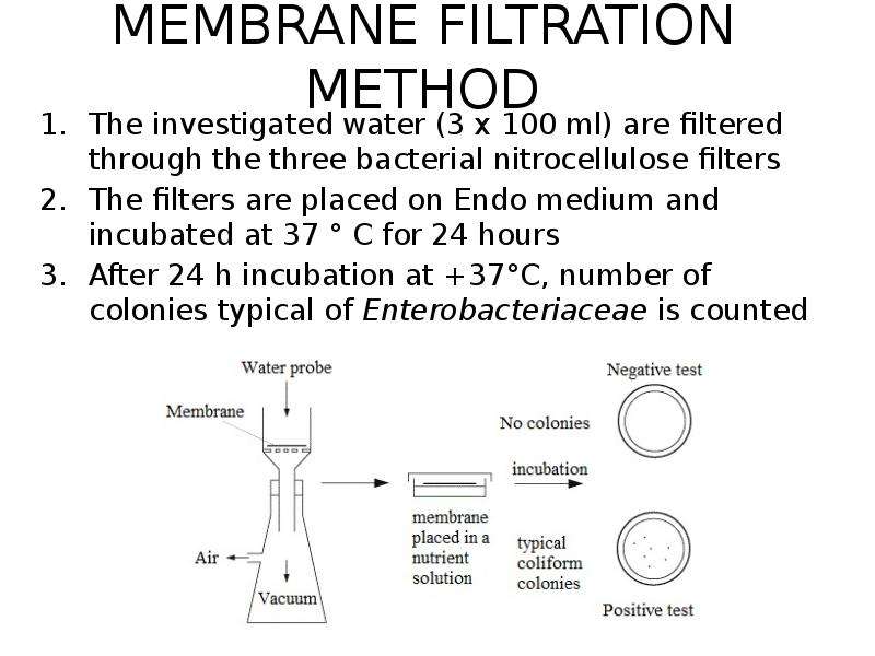
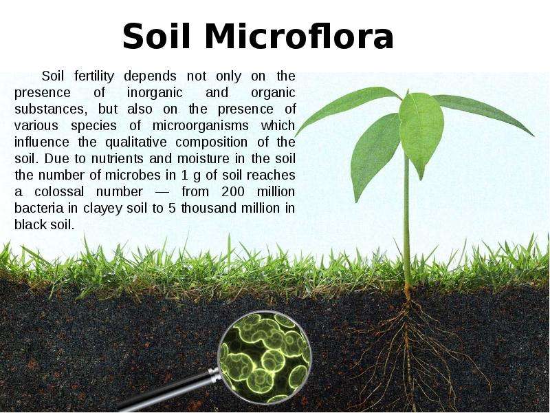
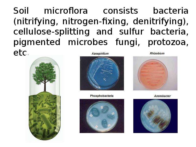
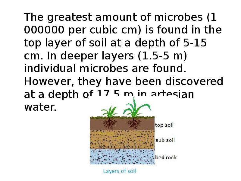
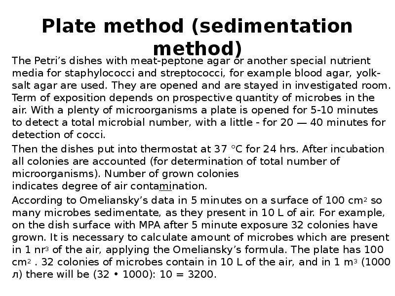
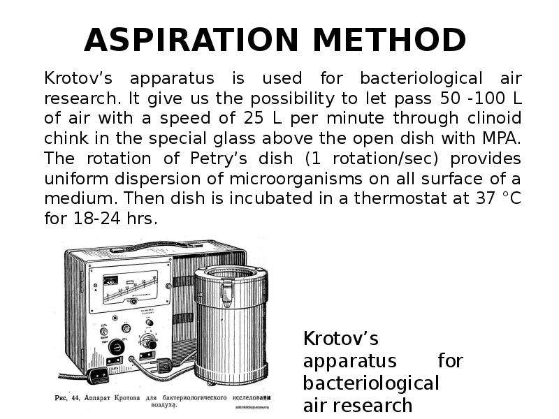
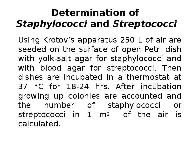
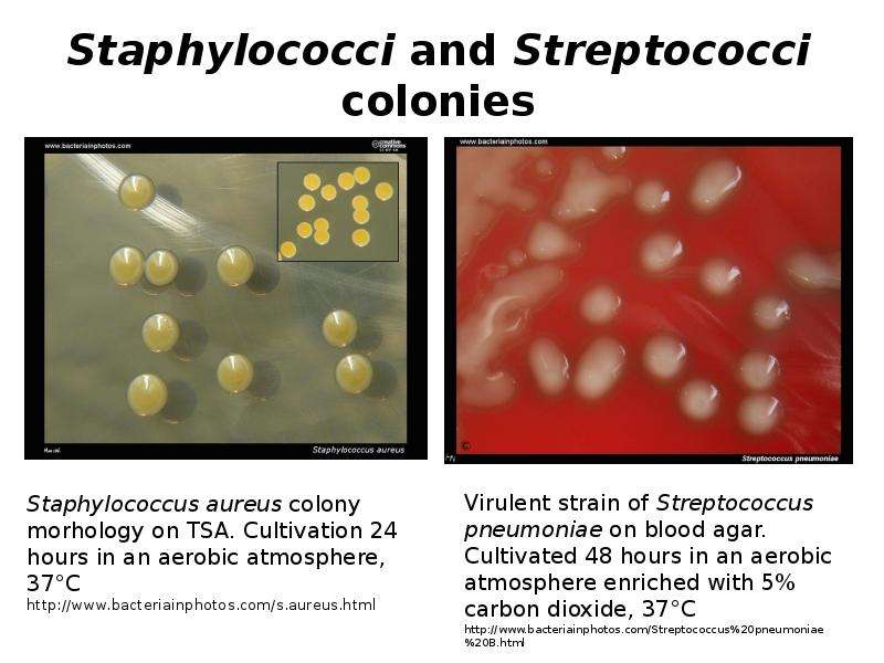
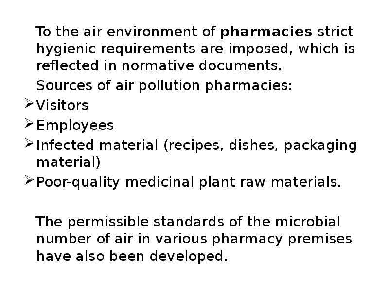
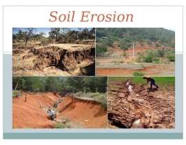
Описание слайда:
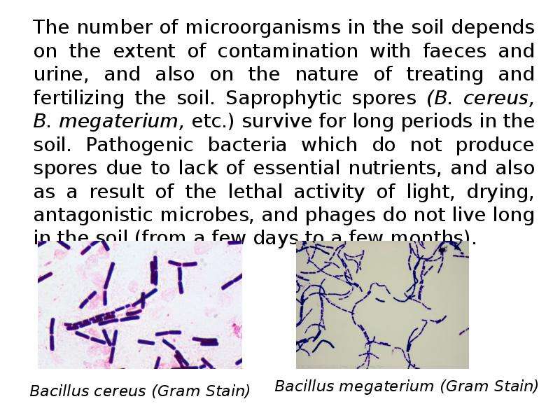
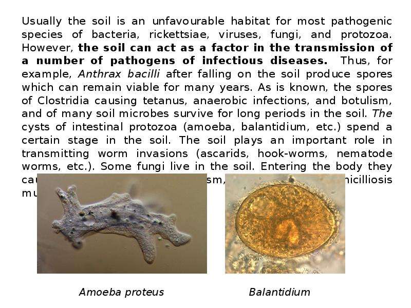
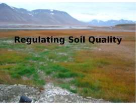
Usually the soil is an unfavourable habitat for most pathogenic species of bacteria, rickettsiae, viruses, fungi, and protozoa. However, the soil can act as a factor in the transmission of a number of pathogens of infectious diseases. Thus, for example, Anthrax bacilli after falling on the soil produce spores which can remain viable for many years. As is known, the spores of Clostridia causing tetanus, anaerobic infections, and botulism, and of many soil microbes survive for long periods in the soil. The cysts of intestinal protozoa (amoeba, balantidium, etc.) spend a certain stage in the soil. The soil plays an important role in transmitting worm invasions (ascarids, hook-worms, nematode worms, etc.). Some fungi live in the soil. Entering the body they cause fusariotoxicosis, ergotism, aspergillosis, penicilliosis mucormycosis, etc. Usually the soil is an unfavourable habitat for most pathogenic species of bacteria, rickettsiae, viruses, fungi, and protozoa. However, the soil can act as a factor in the transmission of a number of pathogens of infectious diseases. Thus, for example, Anthrax bacilli after falling on the soil produce spores which can remain viable for many years. As is known, the spores of Clostridia causing tetanus, anaerobic infections, and botulism, and of many soil microbes survive for long periods in the soil. The cysts of intestinal protozoa (amoeba, balantidium, etc.) spend a certain stage in the soil. The soil plays an important role in transmitting worm invasions (ascarids, hook-worms, nematode worms, etc.). Some fungi live in the soil. Entering the body they cause fusariotoxicosis, ergotism, aspergillosis, penicilliosis mucormycosis, etc.