🗊 Презентация chapt17_apr_lecture2e

Категория: Образование
Нажмите для полного просмотра!
chapt17_apr_lecture2e, слайд №1 chapt17_apr_lecture2e, слайд №2 chapt17_apr_lecture2e, слайд №3 chapt17_apr_lecture2e, слайд №4 chapt17_apr_lecture2e, слайд №5 chapt17_apr_lecture2e, слайд №6 chapt17_apr_lecture2e, слайд №7 chapt17_apr_lecture2e, слайд №8 chapt17_apr_lecture2e, слайд №9 chapt17_apr_lecture2e, слайд №10 chapt17_apr_lecture2e, слайд №11 chapt17_apr_lecture2e, слайд №12 chapt17_apr_lecture2e, слайд №13 chapt17_apr_lecture2e, слайд №14 chapt17_apr_lecture2e, слайд №15 chapt17_apr_lecture2e, слайд №16 chapt17_apr_lecture2e, слайд №17 chapt17_apr_lecture2e, слайд №18 chapt17_apr_lecture2e, слайд №19 chapt17_apr_lecture2e, слайд №20 chapt17_apr_lecture2e, слайд №21 chapt17_apr_lecture2e, слайд №22 chapt17_apr_lecture2e, слайд №23 chapt17_apr_lecture2e, слайд №24 chapt17_apr_lecture2e, слайд №25 chapt17_apr_lecture2e, слайд №26 chapt17_apr_lecture2e, слайд №27 chapt17_apr_lecture2e, слайд №28 chapt17_apr_lecture2e, слайд №29 chapt17_apr_lecture2e, слайд №30 chapt17_apr_lecture2e, слайд №31 chapt17_apr_lecture2e, слайд №32 chapt17_apr_lecture2e, слайд №33 chapt17_apr_lecture2e, слайд №34 chapt17_apr_lecture2e, слайд №35 chapt17_apr_lecture2e, слайд №36 chapt17_apr_lecture2e, слайд №37 chapt17_apr_lecture2e, слайд №38 chapt17_apr_lecture2e, слайд №39 chapt17_apr_lecture2e, слайд №40 chapt17_apr_lecture2e, слайд №41 chapt17_apr_lecture2e, слайд №42 chapt17_apr_lecture2e, слайд №43 chapt17_apr_lecture2e, слайд №44 chapt17_apr_lecture2e, слайд №45 chapt17_apr_lecture2e, слайд №46 chapt17_apr_lecture2e, слайд №47 chapt17_apr_lecture2e, слайд №48 chapt17_apr_lecture2e, слайд №49 chapt17_apr_lecture2e, слайд №50 chapt17_apr_lecture2e, слайд №51 chapt17_apr_lecture2e, слайд №52 chapt17_apr_lecture2e, слайд №53 chapt17_apr_lecture2e, слайд №54 chapt17_apr_lecture2e, слайд №55 chapt17_apr_lecture2e, слайд №56 chapt17_apr_lecture2e, слайд №57 chapt17_apr_lecture2e, слайд №58 chapt17_apr_lecture2e, слайд №59 chapt17_apr_lecture2e, слайд №60 chapt17_apr_lecture2e, слайд №61 chapt17_apr_lecture2e, слайд №62 chapt17_apr_lecture2e, слайд №63 chapt17_apr_lecture2e, слайд №64 chapt17_apr_lecture2e, слайд №65 chapt17_apr_lecture2e, слайд №66 chapt17_apr_lecture2e, слайд №67 chapt17_apr_lecture2e, слайд №68 chapt17_apr_lecture2e, слайд №69 chapt17_apr_lecture2e, слайд №70 chapt17_apr_lecture2e, слайд №71 chapt17_apr_lecture2e, слайд №72 chapt17_apr_lecture2e, слайд №73 chapt17_apr_lecture2e, слайд №74 chapt17_apr_lecture2e, слайд №75 chapt17_apr_lecture2e, слайд №76 chapt17_apr_lecture2e, слайд №77 chapt17_apr_lecture2e, слайд №78 chapt17_apr_lecture2e, слайд №79 chapt17_apr_lecture2e, слайд №80 chapt17_apr_lecture2e, слайд №81 chapt17_apr_lecture2e, слайд №82 chapt17_apr_lecture2e, слайд №83 chapt17_apr_lecture2e, слайд №84 chapt17_apr_lecture2e, слайд №85 chapt17_apr_lecture2e, слайд №86 chapt17_apr_lecture2e, слайд №87 chapt17_apr_lecture2e, слайд №88 chapt17_apr_lecture2e, слайд №89 chapt17_apr_lecture2e, слайд №90 chapt17_apr_lecture2e, слайд №91 chapt17_apr_lecture2e, слайд №92 chapt17_apr_lecture2e, слайд №93 chapt17_apr_lecture2e, слайд №94 chapt17_apr_lecture2e, слайд №95 chapt17_apr_lecture2e, слайд №96 chapt17_apr_lecture2e, слайд №97 chapt17_apr_lecture2e, слайд №98 chapt17_apr_lecture2e, слайд №99 chapt17_apr_lecture2e, слайд №100 chapt17_apr_lecture2e, слайд №101 chapt17_apr_lecture2e, слайд №102 chapt17_apr_lecture2e, слайд №103 chapt17_apr_lecture2e, слайд №104 chapt17_apr_lecture2e, слайд №105 chapt17_apr_lecture2e, слайд №106 chapt17_apr_lecture2e, слайд №107 chapt17_apr_lecture2e, слайд №108 chapt17_apr_lecture2e, слайд №109 chapt17_apr_lecture2e, слайд №110 chapt17_apr_lecture2e, слайд №111 chapt17_apr_lecture2e, слайд №112 chapt17_apr_lecture2e, слайд №113 chapt17_apr_lecture2e, слайд №114 chapt17_apr_lecture2e, слайд №115 chapt17_apr_lecture2e, слайд №116 chapt17_apr_lecture2e, слайд №117 chapt17_apr_lecture2e, слайд №118 chapt17_apr_lecture2e, слайд №119 chapt17_apr_lecture2e, слайд №120 chapt17_apr_lecture2e, слайд №121 chapt17_apr_lecture2e, слайд №122 chapt17_apr_lecture2e, слайд №123 chapt17_apr_lecture2e, слайд №124 chapt17_apr_lecture2e, слайд №125 chapt17_apr_lecture2e, слайд №126 chapt17_apr_lecture2e, слайд №127 chapt17_apr_lecture2e, слайд №128 chapt17_apr_lecture2e, слайд №129 chapt17_apr_lecture2e, слайд №130 chapt17_apr_lecture2e, слайд №131 chapt17_apr_lecture2e, слайд №132 chapt17_apr_lecture2e, слайд №133 chapt17_apr_lecture2e, слайд №134 chapt17_apr_lecture2e, слайд №135 chapt17_apr_lecture2e, слайд №136 chapt17_apr_lecture2e, слайд №137 chapt17_apr_lecture2e, слайд №138 chapt17_apr_lecture2e, слайд №139 chapt17_apr_lecture2e, слайд №140 chapt17_apr_lecture2e, слайд №141 chapt17_apr_lecture2e, слайд №142 chapt17_apr_lecture2e, слайд №143 chapt17_apr_lecture2e, слайд №144 chapt17_apr_lecture2e, слайд №145

Содержание
Вы можете ознакомиться и скачать презентацию на тему chapt17_apr_lecture2e. Доклад-сообщение содержит 145 слайдов. Презентации для любого класса можно скачать бесплатно. Если материал и наш сайт презентаций Mypresentation Вам понравились – поделитесь им с друзьями с помощью социальных кнопок и добавьте в закладки в своем браузере.
Слайды и текст этой презентации


Слайд 1


chapt17_apr_lecture2e, слайд №1
Описание слайда:
Слайд 2


17.1 Introduction to the Endocrine System Compare and contrast the actions of the endocrine system and the nervous system to control body function....
Описание слайда:
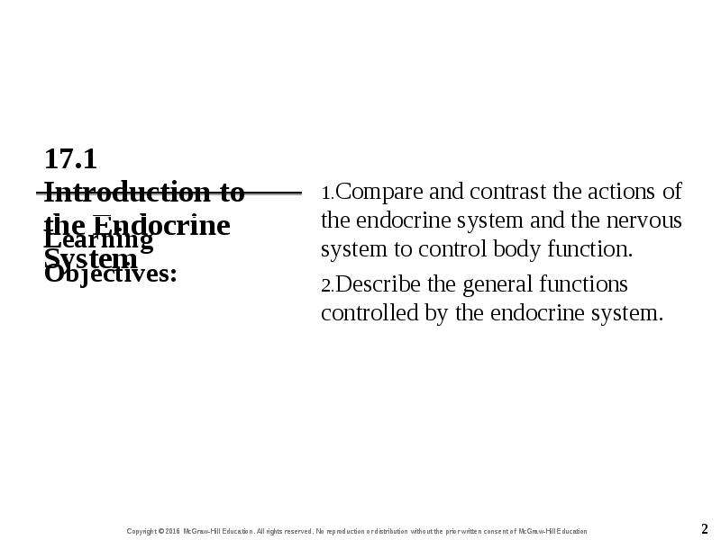
17.1 Introduction to the Endocrine System Compare and contrast the actions of the endocrine system and the nervous system to control body function. Describe the general functions controlled by the endocrine system.

Слайд 3


17.1 Introduction to the Endocrine System Endocrine system Composed of ductless glands that synthesize and secrete hormones Hormones are released...
Описание слайда:
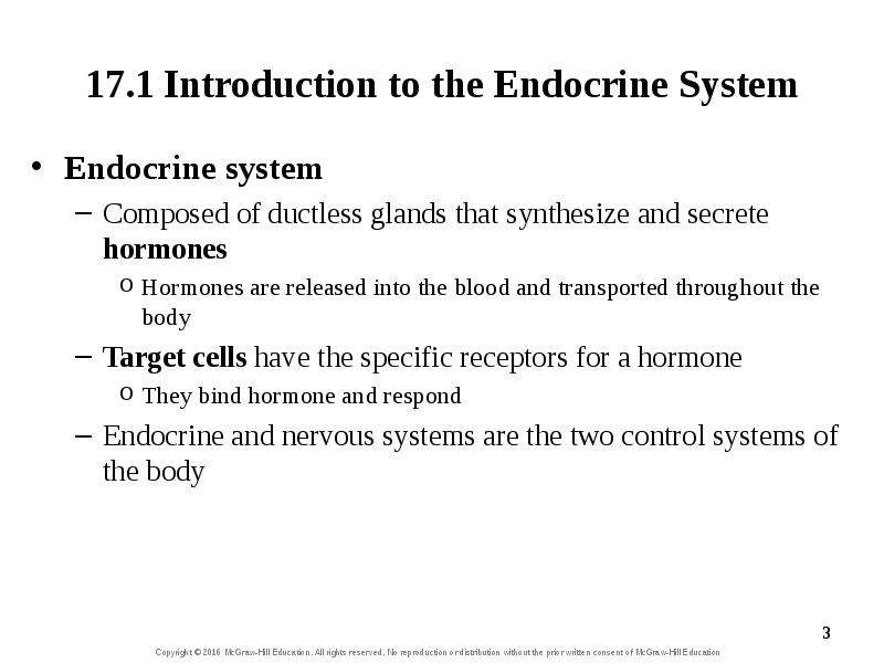
17.1 Introduction to the Endocrine System Endocrine system Composed of ductless glands that synthesize and secrete hormones Hormones are released into the blood and transported throughout the body Target cells have the specific receptors for a hormone They bind hormone and respond Endocrine and nervous systems are the two control systems of the body

Слайд 4


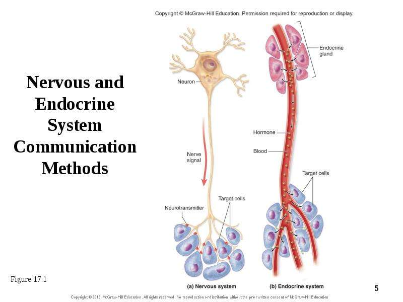
17.1a Comparison of the Two Control Systems Both the endocrine and nervous system Release ligands—chemical messengers Ligands bind to cellular...
Описание слайда:
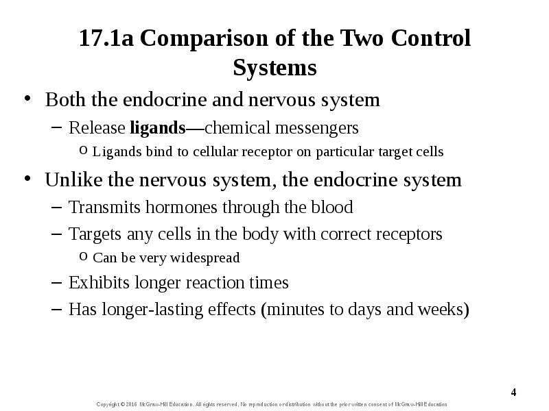
17.1a Comparison of the Two Control Systems Both the endocrine and nervous system Release ligands—chemical messengers Ligands bind to cellular receptor on particular target cells Unlike the nervous system, the endocrine system Transmits hormones through the blood Targets any cells in the body with correct receptors Can be very widespread Exhibits longer reaction times Has longer-lasting effects (minutes to days and weeks)

Слайд 5


Nervous and Endocrine System Communication Methods
Описание слайда:
Nervous and Endocrine System Communication Methods

Слайд 6


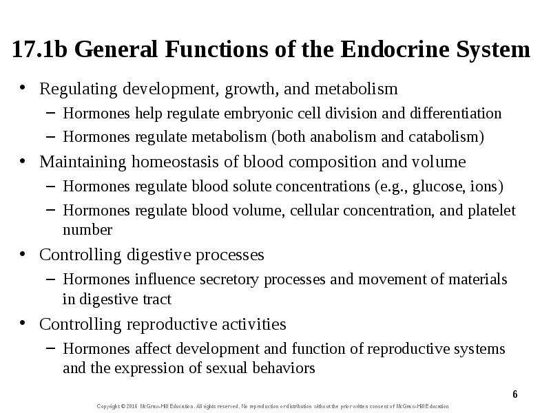
17.1b General Functions of the Endocrine System Regulating development, growth, and metabolism Hormones help regulate embryonic cell division and...
Описание слайда:
17.1b General Functions of the Endocrine System Regulating development, growth, and metabolism Hormones help regulate embryonic cell division and differentiation Hormones regulate metabolism (both anabolism and catabolism) Maintaining homeostasis of blood composition and volume Hormones regulate blood solute concentrations (e.g., glucose, ions) Hormones regulate blood volume, cellular concentration, and platelet number Controlling digestive processes Hormones influence secretory processes and movement of materials in digestive tract Controlling reproductive activities Hormones affect development and function of reproductive systems and the expression of sexual behaviors

Слайд 7


What did you learn? Which control system typically has slower, longer-lasting effects? What general effects can hormones have on the characteristics...
Описание слайда:
What did you learn? Which control system typically has slower, longer-lasting effects? What general effects can hormones have on the characteristics of blood?

Слайд 8


17.2 Endocrine Glands Distinguish between the two types of organization of endocrine cells. Identify the major endocrine glands and their location...
Описание слайда:
17.2 Endocrine Glands Distinguish between the two types of organization of endocrine cells. Identify the major endocrine glands and their location within the body. Explain the three reflex mechanisms for regulating secretion of hormones.

Слайд 9


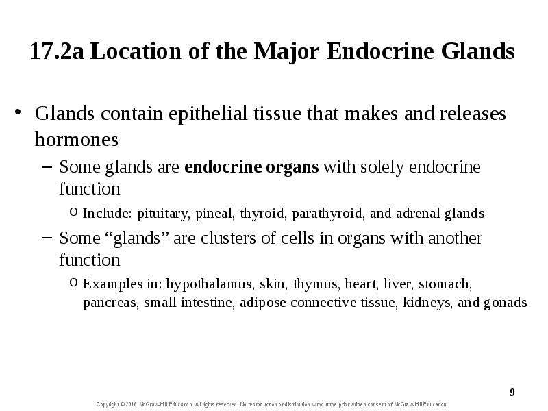
17.2a Location of the Major Endocrine Glands Glands contain epithelial tissue that makes and releases hormones Some glands are endocrine organs with...
Описание слайда:
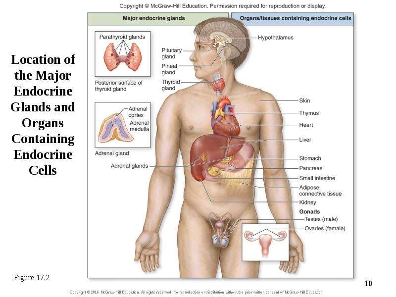
17.2a Location of the Major Endocrine Glands Glands contain epithelial tissue that makes and releases hormones Some glands are endocrine organs with solely endocrine function Include: pituitary, pineal, thyroid, parathyroid, and adrenal glands Some “glands” are clusters of cells in organs with another function Examples in: hypothalamus, skin, thymus, heart, liver, stomach, pancreas, small intestine, adipose connective tissue, kidneys, and gonads

Слайд 10


Location of the Major Endocrine Glands and Organs Containing Endocrine Cells
Описание слайда:
Location of the Major Endocrine Glands and Organs Containing Endocrine Cells

Слайд 11


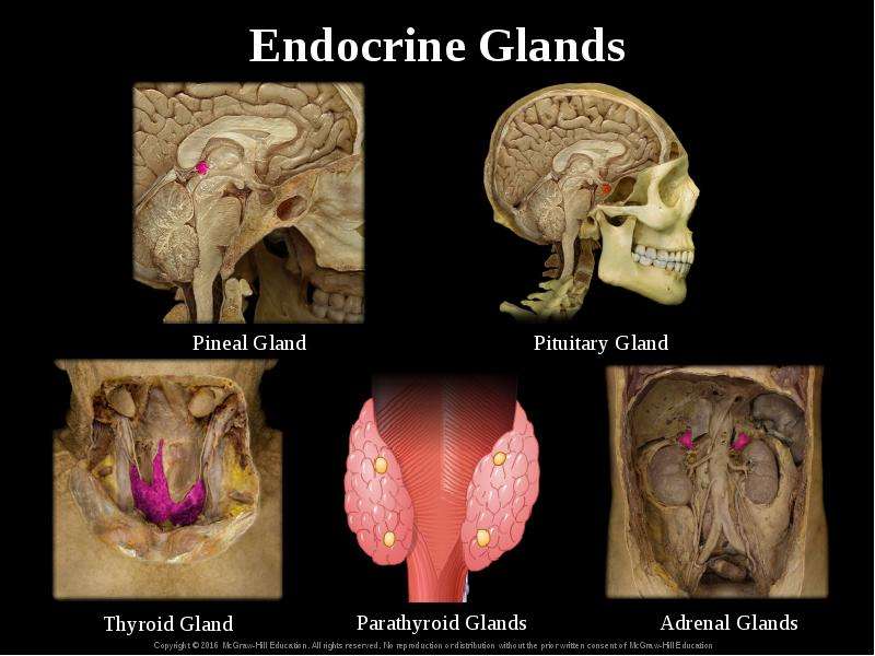
Endocrine Glands
Описание слайда:
Endocrine Glands

Слайд 12


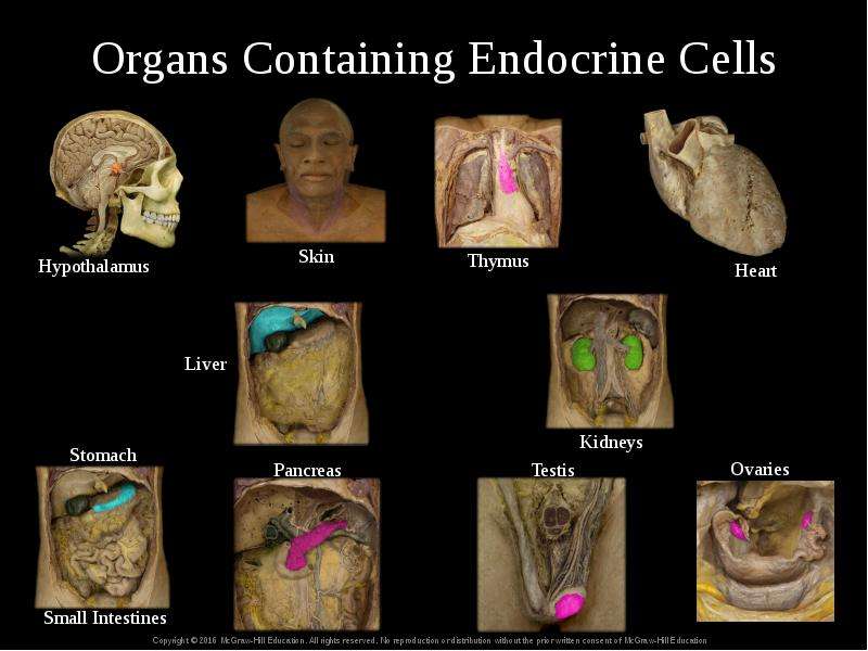
Organs Containing Endocrine Cells
Описание слайда:
Organs Containing Endocrine Cells

Слайд 13


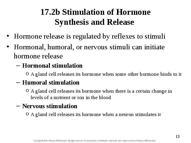
17.2b Stimulation of Hormone Synthesis and Release Hormone release is regulated by reflexes to stimuli Hormonal, humoral, or nervous stimuli can...
Описание слайда:
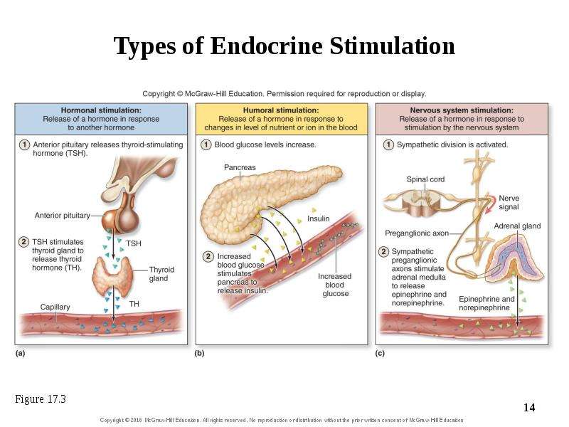
17.2b Stimulation of Hormone Synthesis and Release Hormone release is regulated by reflexes to stimuli Hormonal, humoral, or nervous stimuli can initiate hormone release Hormonal stimulation A gland cell releases its hormone when some other hormone binds to it Humoral stimulation A gland cell releases its hormone when there is a certain change in levels of a nutrient or ion in the blood Nervous stimulation A gland cell releases its hormone when a neuron stimulates it

Слайд 14


Types of Endocrine Stimulation
Описание слайда:
Types of Endocrine Stimulation

Слайд 15


What did you learn? Is the entire pancreas an endocrine organ? Parathyroid hormone is secreted when blood calcium levels drop too low. What sort of...
Описание слайда:
What did you learn? Is the entire pancreas an endocrine organ? Parathyroid hormone is secreted when blood calcium levels drop too low. What sort of stimulation is this?

Слайд 16


17.3 Hormones Name the three structural categories of circulating hormones, and give examples within each category. Distinguish the hormones that are...
Описание слайда:
17.3 Hormones Name the three structural categories of circulating hormones, and give examples within each category. Distinguish the hormones that are lipid-soluble from those that are water-soluble. Describe the general structure, formation, and function of local hormones. Compare autocrine and paracrine signaling that occurs through local hormones.

Слайд 17


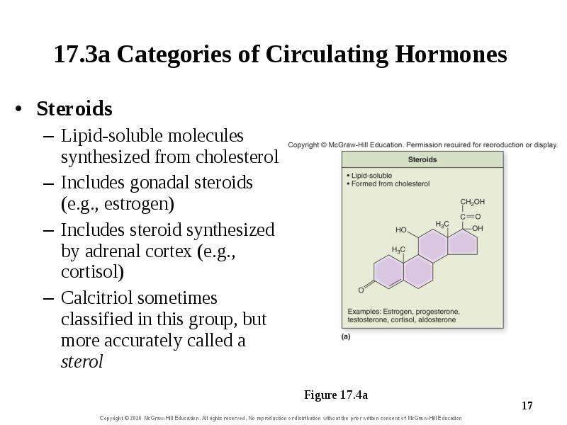
17.3a Categories of Circulating Hormones Steroids Lipid-soluble molecules synthesized from cholesterol Includes gonadal steroids (e.g., estrogen)...
Описание слайда:
17.3a Categories of Circulating Hormones Steroids Lipid-soluble molecules synthesized from cholesterol Includes gonadal steroids (e.g., estrogen) Includes steroid synthesized by adrenal cortex (e.g., cortisol) Calcitriol sometimes classified in this group, but more accurately called a sterol

Слайд 18


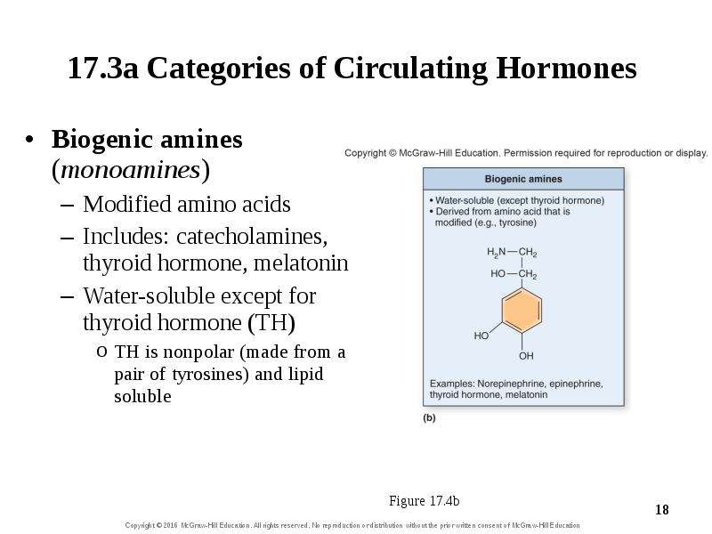
17.3a Categories of Circulating Hormones Biogenic amines (monoamines) Modified amino acids Includes: catecholamines, thyroid hormone, melatonin...
Описание слайда:
17.3a Categories of Circulating Hormones Biogenic amines (monoamines) Modified amino acids Includes: catecholamines, thyroid hormone, melatonin Water-soluble except for thyroid hormone (TH) TH is nonpolar (made from a pair of tyrosines) and lipid soluble

Слайд 19


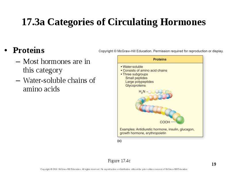
17.3a Categories of Circulating Hormones Proteins Most hormones are in this category Water-soluble chains of amino acids
Описание слайда:
17.3a Categories of Circulating Hormones Proteins Most hormones are in this category Water-soluble chains of amino acids

Слайд 20


17.3b Local Hormones Local hormones Signaling molecules that don’t circulate in blood Some biologists don’t consider them “hormones” They bind to...
Описание слайда:
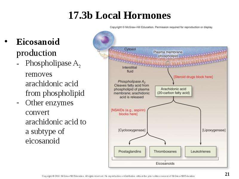
17.3b Local Hormones Local hormones Signaling molecules that don’t circulate in blood Some biologists don’t consider them “hormones” They bind to neighboring cells or the cells that release them Eicosanoids: a type of local hormone formed from fatty acids within phospholipid bilayer of membrane Synthesized through an enzymatic cascade

Слайд 21


chapt17_apr_lecture2e, слайд №21
Описание слайда:

Слайд 22


17.3b Local Hormones Eicosanoid effects Autocrine stimulation Effects on the same cell where messenger was formed Paracrine stimulation Effects on...
Описание слайда:
17.3b Local Hormones Eicosanoid effects Autocrine stimulation Effects on the same cell where messenger was formed Paracrine stimulation Effects on neighboring cells Prostaglandins are eicosanoids Stimulate pain and inflammatory responses Aspirin and other nonsteroidal anti-inflammatory drugs block prostaglandin formation

Слайд 23


What did you learn? Insulin is made up of a chain of amino acids. What class of hormone is it? Is it water soluble or lipid soluble? How are...
Описание слайда:
What did you learn? Insulin is made up of a chain of amino acids. What class of hormone is it? Is it water soluble or lipid soluble? How are prostaglandins synthesized?

Слайд 24


17.4 Hormone Transport Compare the transport of lipid-soluble hormones with that of water-soluble hormones. Describe the two primary factors that...
Описание слайда:
17.4 Hormone Transport Compare the transport of lipid-soluble hormones with that of water-soluble hormones. Describe the two primary factors that affect the concentration level of a circulating hormone. Explain what is meant by the half-life of a hormone.

Слайд 25


17.4a Transport in the Blood Lipid-soluble hormones use carrier molecules Do not dissolve readily in blood Carriers are water-soluble proteins made...
Описание слайда:
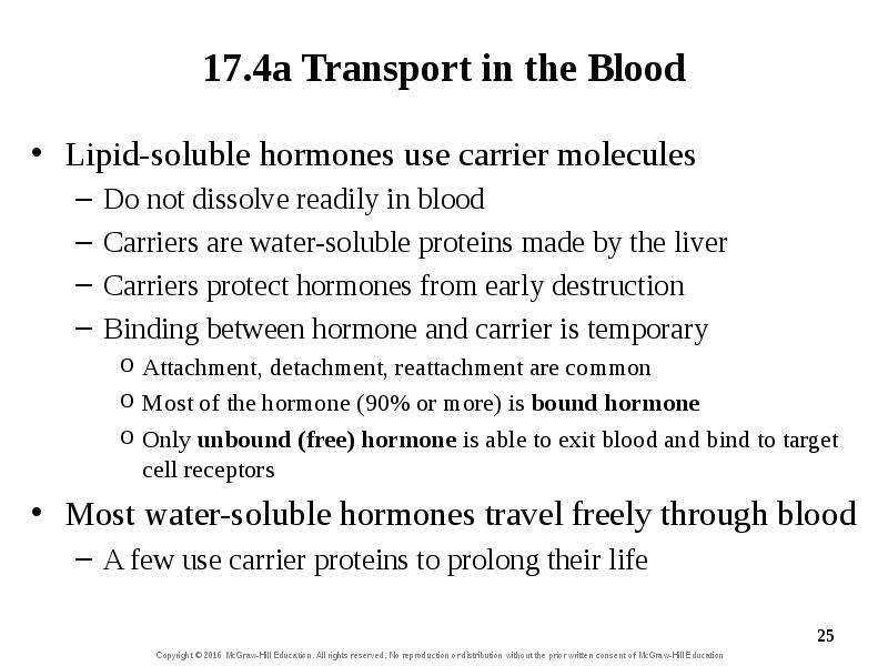
17.4a Transport in the Blood Lipid-soluble hormones use carrier molecules Do not dissolve readily in blood Carriers are water-soluble proteins made by the liver Carriers protect hormones from early destruction Binding between hormone and carrier is temporary Attachment, detachment, reattachment are common Most of the hormone (90% or more) is bound hormone Only unbound (free) hormone is able to exit blood and bind to target cell receptors Most water-soluble hormones travel freely through blood A few use carrier proteins to prolong their life

Слайд 26


17.4b Levels of Circulating Hormone A hormone’s blood concentration depends on how fast it is synthesized and eliminated Hormone synthesis is done by...
Описание слайда:
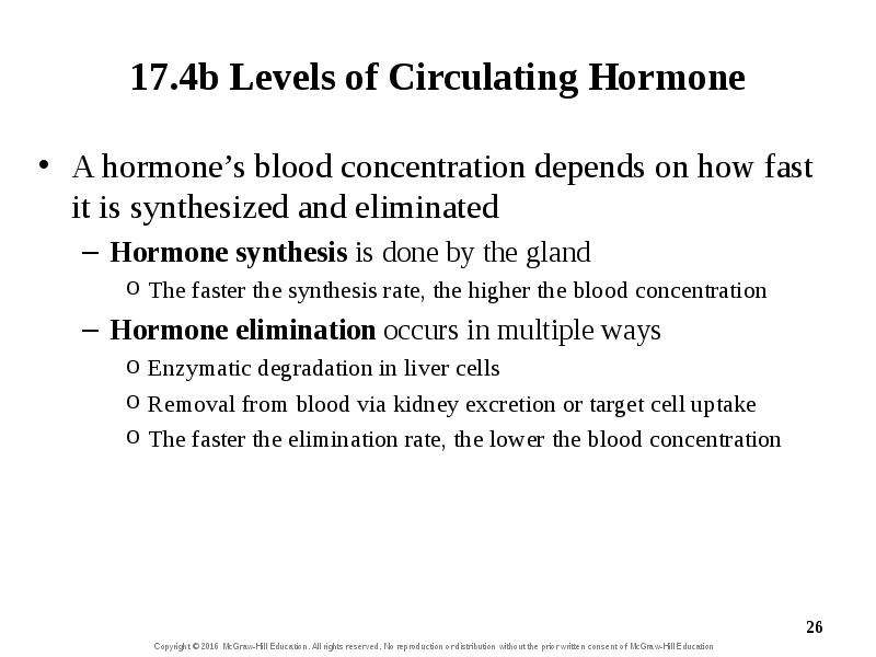
17.4b Levels of Circulating Hormone A hormone’s blood concentration depends on how fast it is synthesized and eliminated Hormone synthesis is done by the gland The faster the synthesis rate, the higher the blood concentration Hormone elimination occurs in multiple ways Enzymatic degradation in liver cells Removal from blood via kidney excretion or target cell uptake The faster the elimination rate, the lower the blood concentration

Слайд 27


17.4b Levels of Circulating Hormone Half-Life—time necessary to reduce a hormone’s concentration to half of its original level Depends on how...
Описание слайда:
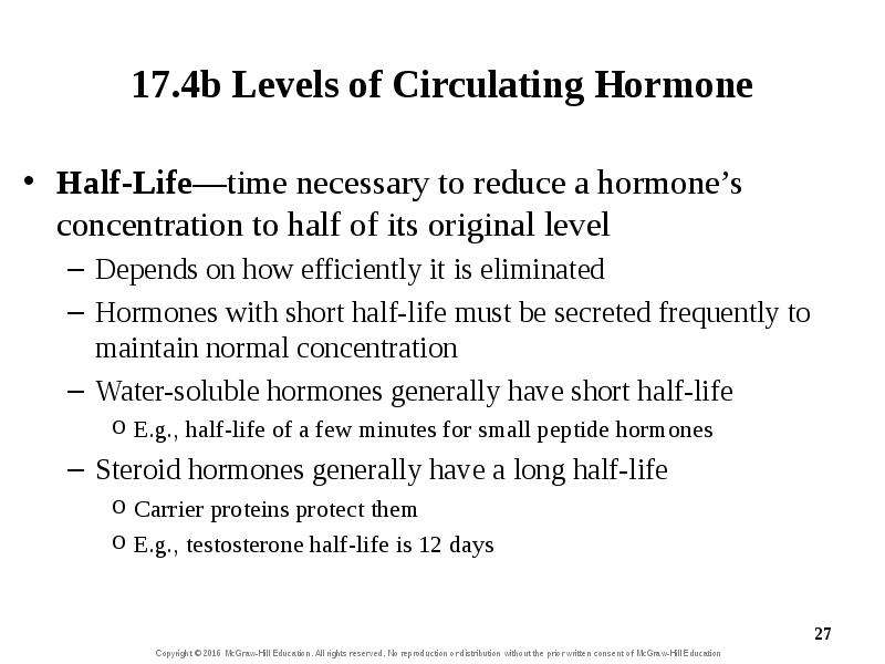
17.4b Levels of Circulating Hormone Half-Life—time necessary to reduce a hormone’s concentration to half of its original level Depends on how efficiently it is eliminated Hormones with short half-life must be secreted frequently to maintain normal concentration Water-soluble hormones generally have short half-life E.g., half-life of a few minutes for small peptide hormones Steroid hormones generally have a long half-life Carrier proteins protect them E.g., testosterone half-life is 12 days

Слайд 28


What did you learn? If hormone X and hormone Y had the same rate of synthesis, but X’s elimination rate was faster, which would be at a higher level...
Описание слайда:
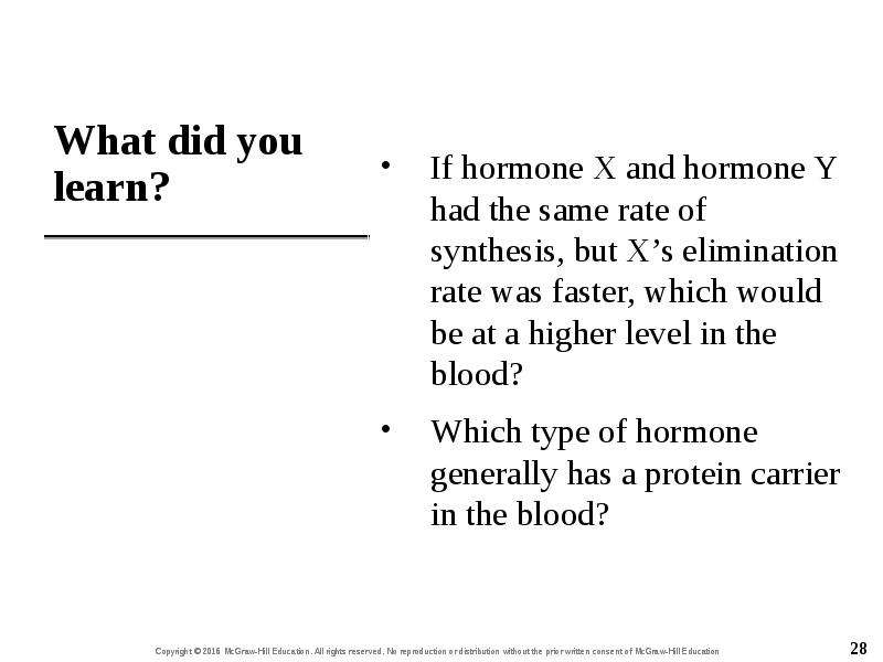
What did you learn? If hormone X and hormone Y had the same rate of synthesis, but X’s elimination rate was faster, which would be at a higher level in the blood? Which type of hormone generally has a protein carrier in the blood?

Слайд 29


17.5 Target Cells: Interactions with Hormones Describe how lipid-soluble hormones reach their target cell receptors and the type of cellular change...
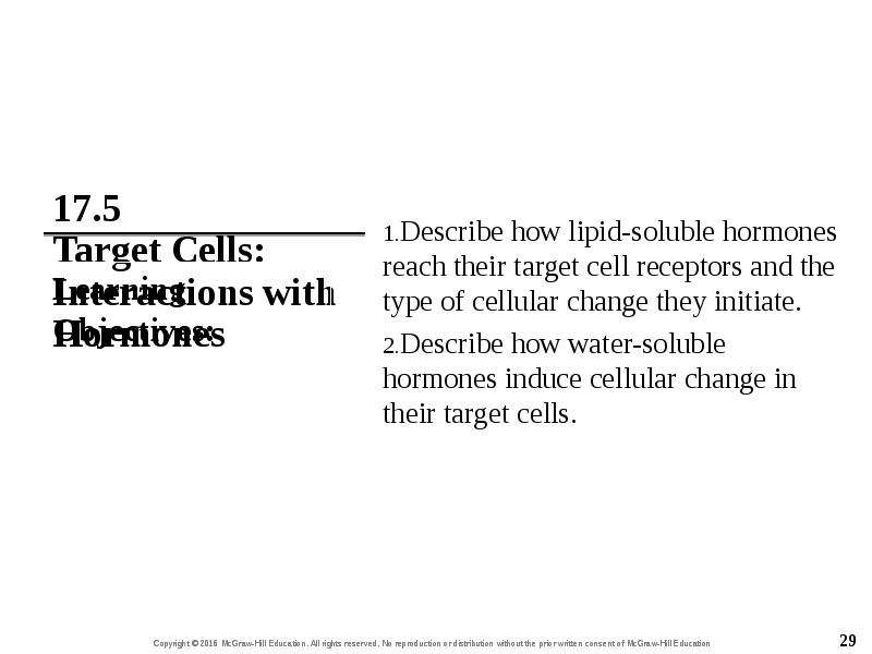
Описание слайда:
17.5 Target Cells: Interactions with Hormones Describe how lipid-soluble hormones reach their target cell receptors and the type of cellular change they initiate. Describe how water-soluble hormones induce cellular change in their target cells.

Слайд 30


17.5a Lipid-Soluble Hormones Lipid-soluble hormones can diffuse across target cell membrane Such hormones are small, nonpolar, and lipophilic Their...
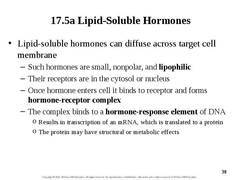
Описание слайда:
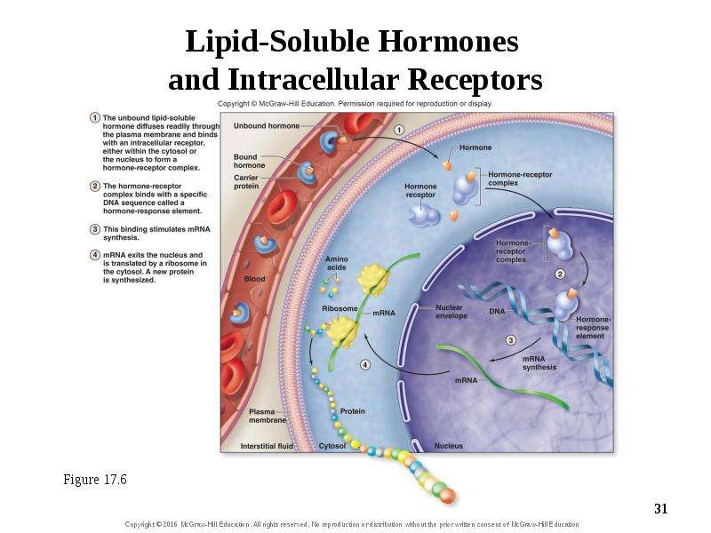
17.5a Lipid-Soluble Hormones Lipid-soluble hormones can diffuse across target cell membrane Such hormones are small, nonpolar, and lipophilic Their receptors are in the cytosol or nucleus Once hormone enters cell it binds to receptor and forms hormone-receptor complex The complex binds to a hormone-response element of DNA Results in transcription of an mRNA, which is translated to a protein The protein may have structural or metabolic effects

Слайд 31


Figure 17.6
Описание слайда:
Figure 17.6

Слайд 32


17.5b Water-Soluble Hormones Water-soluble hormones use membrane receptors Such hormones are polar and can’t diffuse through membrane Signal...
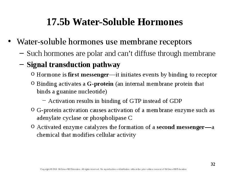
Описание слайда:
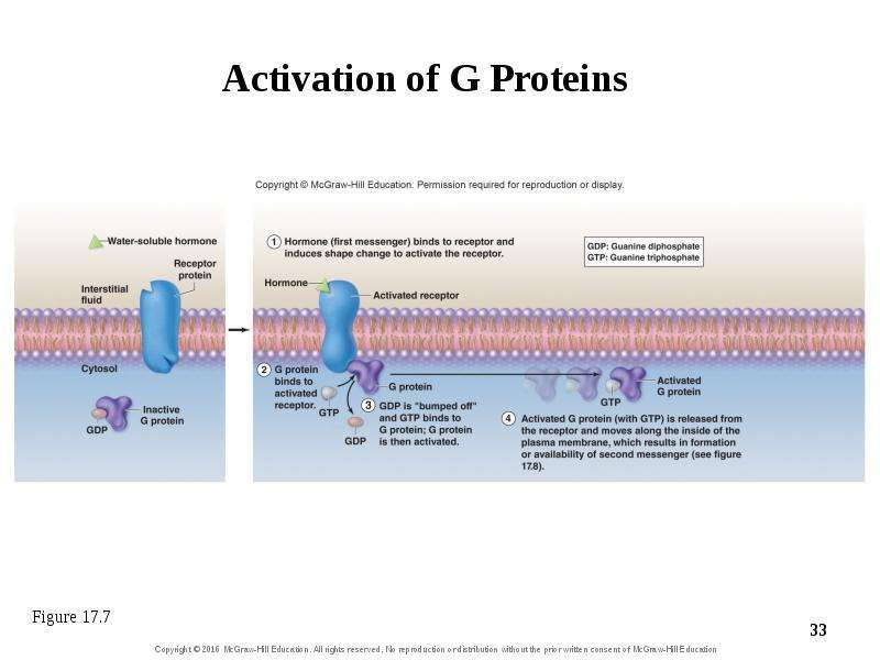
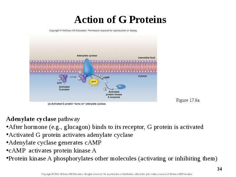
17.5b Water-Soluble Hormones Water-soluble hormones use membrane receptors Such hormones are polar and can’t diffuse through membrane Signal transduction pathway Hormone is first messenger—it initiates events by binding to receptor Binding activates a G-protein (an internal membrane protein that binds a guanine nucleotide) Activation results in binding of GTP instead of GDP G-protein activation causes activation of a membrane enzyme such as adenylate cyclase or phospholipase C Activated enzyme catalyzes the formation of a second messenger—a chemical that modifies cellular activity

Слайд 33


Activation of G Proteins
Описание слайда:
Activation of G Proteins

Слайд 34


chapt17_apr_lecture2e, слайд №34
Описание слайда:

Слайд 35


17.5b Water-Soluble Hormones Phospholipase C pathway After hormone (e.g., epinephrine) binds to its receptor, G protein is activated Activated G...
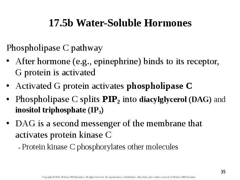
Описание слайда:
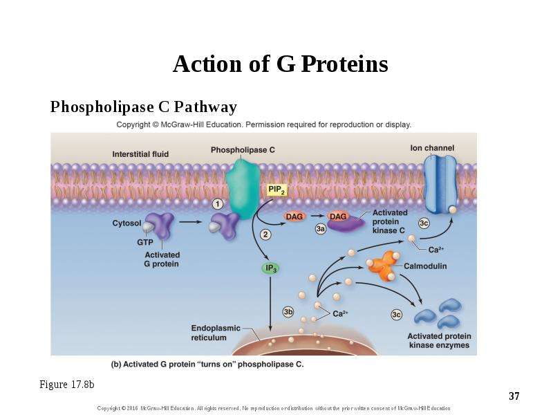
17.5b Water-Soluble Hormones Phospholipase C pathway After hormone (e.g., epinephrine) binds to its receptor, G protein is activated Activated G protein activates phospholipase C Phospholipase C splits PIP2 into diacylglycerol (DAG) and inositol triphosphate (IP3) DAG is a second messenger of the membrane that activates protein kinase C - Protein kinase C phosphorylates other molecules

Слайд 36


17.5b Water-Soluble Hormones Phospholipase C pathway (continued) IP3 is a second messenger that leaves the membrane and causes an increase in the...
Описание слайда:
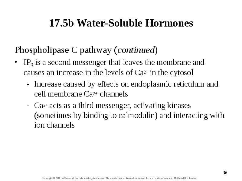
17.5b Water-Soluble Hormones Phospholipase C pathway (continued) IP3 is a second messenger that leaves the membrane and causes an increase in the levels of Ca2+ in the cytosol Increase caused by effects on endoplasmic reticulum and cell membrane Ca2+ channels Ca2+ acts as a third messenger, activating kinases (sometimes by binding to calmodulin) and interacting with ion channels

Слайд 37


chapt17_apr_lecture2e, слайд №37
Описание слайда:

Слайд 38


17.5b Water-Soluble Hormones Action of water-soluble hormones Multiple results possible with different signal transduction pathways Enzymes can be...
Описание слайда:
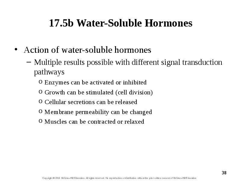
17.5b Water-Soluble Hormones Action of water-soluble hormones Multiple results possible with different signal transduction pathways Enzymes can be activated or inhibited Growth can be stimulated (cell division) Cellular secretions can be released Membrane permeability can be changed Muscles can be contracted or relaxed

Слайд 39


17.5b Water-Soluble Hormones Action of water-soluble hormones (continued) E.g., glucagon released from pancreas when blood sugar is low Binds to...
Описание слайда:
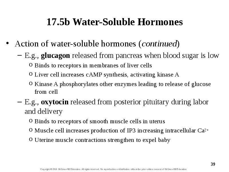
17.5b Water-Soluble Hormones Action of water-soluble hormones (continued) E.g., glucagon released from pancreas when blood sugar is low Binds to receptors in membranes of liver cells Liver cell increases cAMP synthesis, activating kinase A Kinase A phosphorylates other enzymes leading to release of glucose from cell E.g., oxytocin released from posterior pituitary during labor and delivery Binds to receptors of smooth muscle cells in uterus Muscle cell increases production of IP3 increasing intracellular Ca2+ Uterine muscle contractions strengthen to expel baby

Слайд 40


17.5b Water-Soluble Hormones Intracellular enzyme cascade and response amplification Signaling pathway advantages Signal is amplified at each...
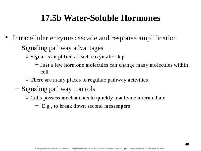
Описание слайда:
17.5b Water-Soluble Hormones Intracellular enzyme cascade and response amplification Signaling pathway advantages Signal is amplified at each enzymatic step Just a few hormone molecules can change many molecules within cell There are many places to regulate pathway activities Signaling pathway controls Cells possess mechanisms to quickly inactivate intermediate E.g., to break down second messengers

Слайд 41


What did you learn? Where are target cell receptors for lipophilic hormones located? What is protein kinase A, and what role does it have in a signal...
Описание слайда:
What did you learn? Where are target cell receptors for lipophilic hormones located? What is protein kinase A, and what role does it have in a signal pathway? Where does DAG come from, and what function does it serve?

Слайд 42


17.6 Target Cells: Degree of Cellular Response Describe the conditions that influence the number of receptors available for a specific hormone....
Описание слайда:
17.6 Target Cells: Degree of Cellular Response Describe the conditions that influence the number of receptors available for a specific hormone. Define up-regulation and down-regulation. Compare and contrast the three types of hormone interactions.

Слайд 43


17.6 Target Cells: Degree of Cellular Response A cell’s response to a hormone varies with Its number of receptors for the hormone Its simultaneous...
Описание слайда:
17.6 Target Cells: Degree of Cellular Response A cell’s response to a hormone varies with Its number of receptors for the hormone Its simultaneous response to other hormones

Слайд 44


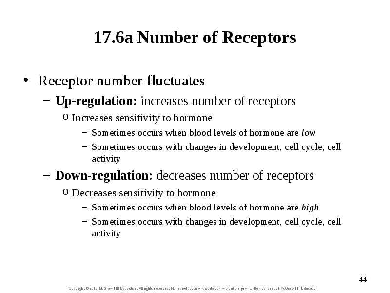
17.6a Number of Receptors Receptor number fluctuates Up-regulation: increases number of receptors Increases sensitivity to hormone Sometimes occurs...
Описание слайда:
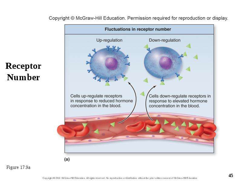
17.6a Number of Receptors Receptor number fluctuates Up-regulation: increases number of receptors Increases sensitivity to hormone Sometimes occurs when blood levels of hormone are low Sometimes occurs with changes in development, cell cycle, cell activity Down-regulation: decreases number of receptors Decreases sensitivity to hormone Sometimes occurs when blood levels of hormone are high Sometimes occurs with changes in development, cell cycle, cell activity

Слайд 45


Receptor Number
Описание слайда:
Receptor Number

Слайд 46


17.6b Receptor Interactions Different hormones can simultaneously bind to a cell Synergistic interactions One hormone reinforces activity of another...
Описание слайда:
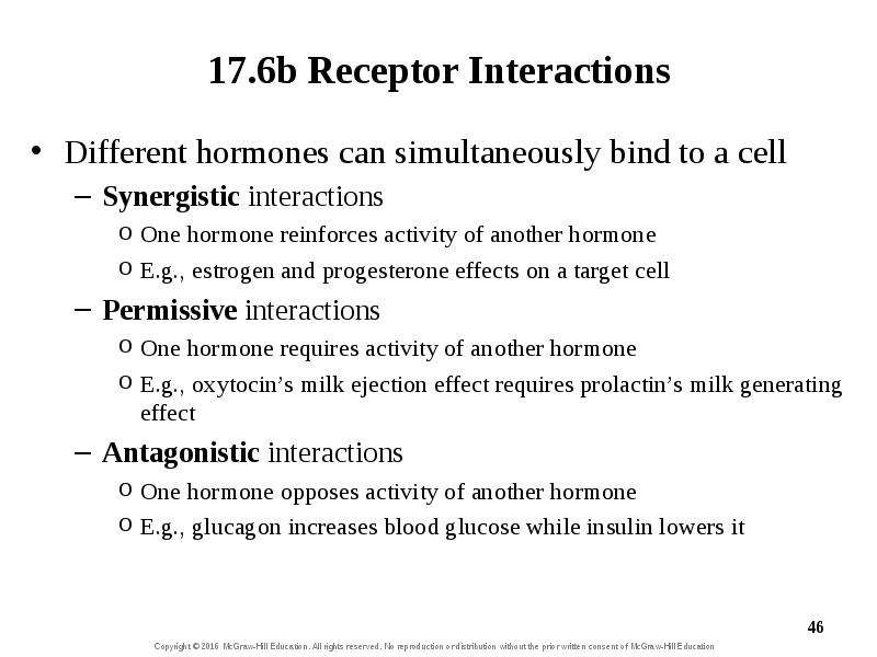
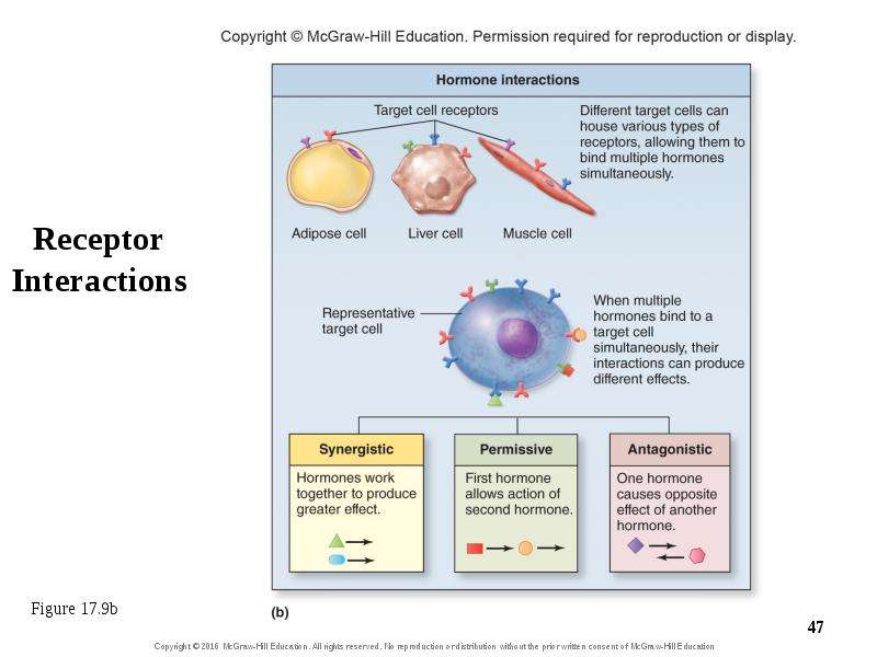
17.6b Receptor Interactions Different hormones can simultaneously bind to a cell Synergistic interactions One hormone reinforces activity of another hormone E.g., estrogen and progesterone effects on a target cell Permissive interactions One hormone requires activity of another hormone E.g., oxytocin’s milk ejection effect requires prolactin’s milk generating effect Antagonistic interactions One hormone opposes activity of another hormone E.g., glucagon increases blood glucose while insulin lowers it

Слайд 47


Receptor Interactions
Описание слайда:
Receptor Interactions

Слайд 48


What did you learn? If someone were to take a large dose of artificial hormone, how might target cells respond to maintain a normal level of...
Описание слайда:
What did you learn? If someone were to take a large dose of artificial hormone, how might target cells respond to maintain a normal level of response? What type of interaction occurs when a target cell has receptors for two hormones causing opposing effects?

Слайд 49


17.7 The Hypothalamus and the Pituitary Gland Describe the anatomic relationship of the hypothalamus and the pituitary gland. Identify the specific...
Описание слайда:
17.7 The Hypothalamus and the Pituitary Gland Describe the anatomic relationship of the hypothalamus and the pituitary gland. Identify the specific structures associated with the posterior pituitary and the anterior pituitary. Identify the two hormones released from the posterior pituitary, and describe how the hypothalamus controls their release. List the hormones released from the hypothalamus that control the anterior pituitary. Explain how the hypothalamus controls the release of hormones from the anterior pituitary and the general function of each.

Слайд 50


17.7a Anatomic Relationship of the Hypothalamus and the Pituitary Gland Hypothalamus controls pituitary, which controls thyroid, adrenal, liver,...
Описание слайда:
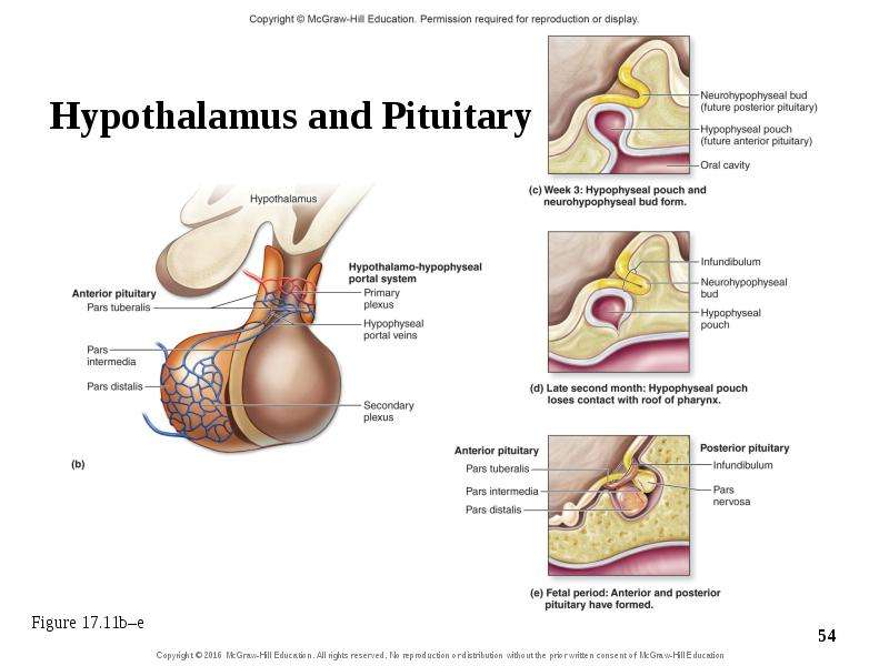
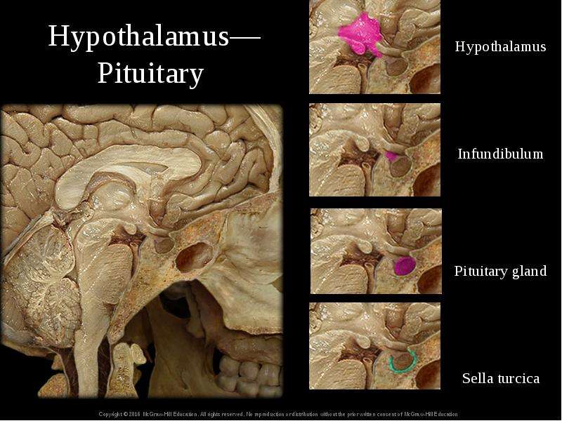
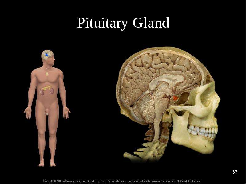
17.7a Anatomic Relationship of the Hypothalamus and the Pituitary Gland Hypothalamus controls pituitary, which controls thyroid, adrenal, liver, testes, ovaries Pituitary gland (hypophysis) Lies inferior to hypothalamus in sella turcica of sphenoid bone Pea sized Connected to hypothalamus by infundibulum (stalk) Partitioned into anterior and posterior pituitary (lobes)

Слайд 51


Hypothalamus and Pituitary
Описание слайда:
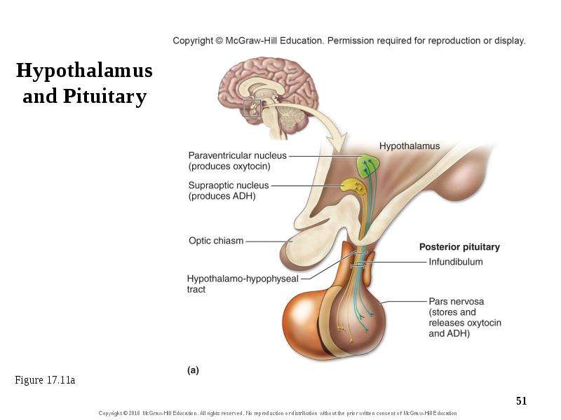
Hypothalamus and Pituitary

Слайд 52


17.7a Anatomic Relationship of the Hypothalamus and the Pituitary Gland Posterior pituitary (neurohypophysis) Smaller, neural part of pituitary gland...
Описание слайда:
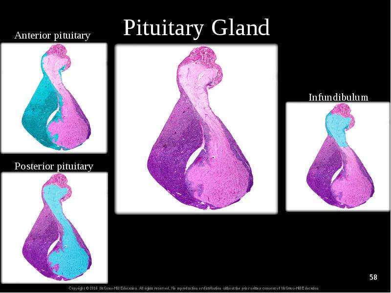
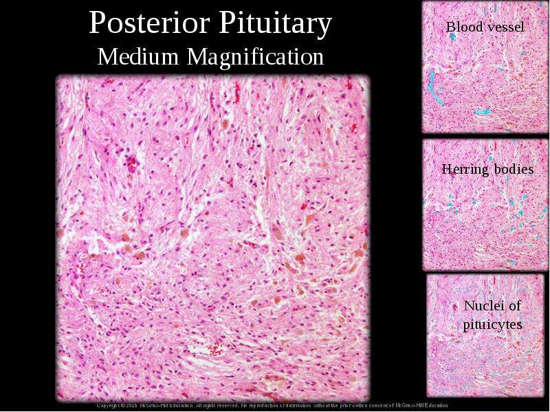
17.7a Anatomic Relationship of the Hypothalamus and the Pituitary Gland Posterior pituitary (neurohypophysis) Smaller, neural part of pituitary gland Develops as a bud from the developing hypothalamus Composed of pars nervosa (lobe) and infundibulum Hypothalamic neurons project through infundibulum and release hormones in pars nervosa Somas in paraventricular nucleus and suprapotic nucleus Axons in hypothalmo-hypophyseal tract of infundibulum Synaptic knobs in pars nervosa

Слайд 53


17.7a Anatomic Relationship of the Hypothalamus and the Pituitary Gland Anterior pituitary (adenohypophysis) Larger, glandular part of pituitary...
Описание слайда:
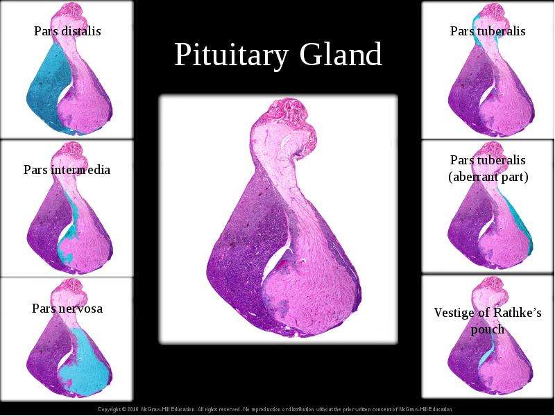
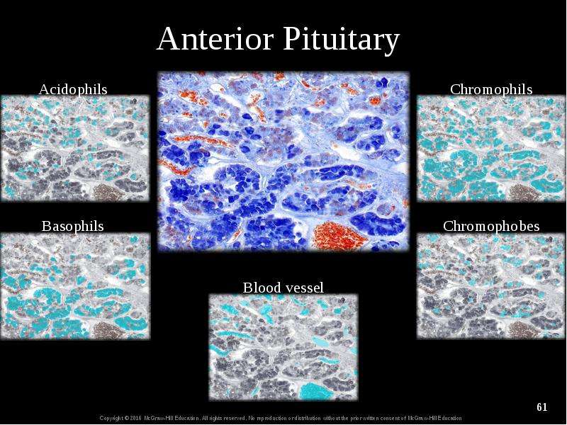
17.7a Anatomic Relationship of the Hypothalamus and the Pituitary Gland Anterior pituitary (adenohypophysis) Larger, glandular part of pituitary Develops from ectoderm of oral cavity Partitioned into three areas: Pars distalis, large anterior rounded portion Pars tuberalis, thin wrapping around infundibulum Pars intermedia, scant region between the other two areas

Слайд 54


chapt17_apr_lecture2e, слайд №54
Описание слайда:

Слайд 55


17.7a Anatomic Relationship of the Hypothalamus and the Pituitary Gland Anterior pituitary (continued) Hypothalamo-hypophyseal portal system of blood...
Описание слайда:
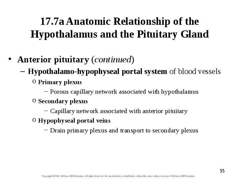
17.7a Anatomic Relationship of the Hypothalamus and the Pituitary Gland Anterior pituitary (continued) Hypothalamo-hypophyseal portal system of blood vessels Primary plexus Porous capillary network associated with hypothalamus Secondary plexus Capillary network associated with anterior pituitary Hypophyseal portal veins Drain primary plexus and transport to secondary plexus

Слайд 56


Hypothalamus—Pituitary
Описание слайда:
Hypothalamus—Pituitary

Слайд 57


Pituitary Gland
Описание слайда:
Pituitary Gland

Слайд 58


Pituitary Gland
Описание слайда:
Pituitary Gland

Слайд 59


Posterior Pituitary Medium Magnification
Описание слайда:
Posterior Pituitary Medium Magnification

Слайд 60


Pituitary Gland
Описание слайда:
Pituitary Gland

Слайд 61


Anterior Pituitary
Описание слайда:
Anterior Pituitary

Слайд 62


17.7b Interactions Between the Hypothalamus and the Posterior Pituitary Gland Posterior pituitary is storage and release site for oxytocin (OT) and...
Описание слайда:
17.7b Interactions Between the Hypothalamus and the Posterior Pituitary Gland Posterior pituitary is storage and release site for oxytocin (OT) and antidiuretic hormone (ADH) Hormones made in hypothalamus by neurosecretory cells Packed in secretory vesicles, transported by fast axonal transport Released from synaptic knobs into blood when neurons fire impulses Oxytocin Made in paraventricular nucleus Functions: uterine contraction, milk ejection , emotional bonding Antidiuretic hormone (vasopressin) Made in supraoptic nucleus Functions: decrease urine production, stimulate thirst, constrict blood vessels

Слайд 63


17.7c Interactions Between the Hypothalamus and the Anterior Pituitary Gland Hypothalamus hormonally stimulates anterior pituitary to release its...
Описание слайда:
17.7c Interactions Between the Hypothalamus and the Anterior Pituitary Gland Hypothalamus hormonally stimulates anterior pituitary to release its hormones Hypothalamus secretes regulatory hormones Travel via portal blood vessels to pituitary Anterior pituitary secretes hormones into general circulation

Слайд 64


17.7c Interactions Between the Hypothalamus and the Anterior Pituitary Gland Regulatory hormones of the hypothalamus Releasing hormones Increase...
Описание слайда:
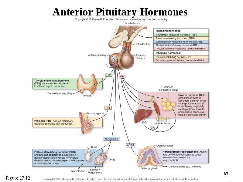
17.7c Interactions Between the Hypothalamus and the Anterior Pituitary Gland Regulatory hormones of the hypothalamus Releasing hormones Increase secretion of anterior pituitary hormones Include: thyrotropin-releasing hormone (TRH), prolactin-releasing hormone (PRH), gonadotropin-releasing hormone (GnRH), corticotropin-releasing hormone (CRH), and growth hormone-releasing hormone (GHRH). Inhibiting hormones Decrease secretion of anterior pituitary hormones Include: prolactin-inhibiting hormone (PIH) and growth-inhibiting hormone (GIH)

Слайд 65


17.7c Interactions Between the Hypothalamus and the Anterior Pituitary Gland Anterior pituitary—tropic hormones and prolactin Thyroid stimulating...
Описание слайда:
17.7c Interactions Between the Hypothalamus and the Anterior Pituitary Gland Anterior pituitary—tropic hormones and prolactin Thyroid stimulating hormone (TSH) Release triggered by TRH from hypothalamus Causes release of thyroid hormone (TH) from thyroid gland Prolactin (PRL) Release triggered by PRH, inhibited by PIH from hypothalamus Causes milk production, mammary gland growth in females Adrenocorticotropic hormone (ACTH; corticotropin) Release triggered by CRH from hypothalamus Causes release of corticosteroids by adrenal cortex

Слайд 66


17.7c Interactions Between the Hypothalamus and the Anterior Pituitary Gland Anterior pituitary—tropic hormones and prolactin (continued)...
Описание слайда:
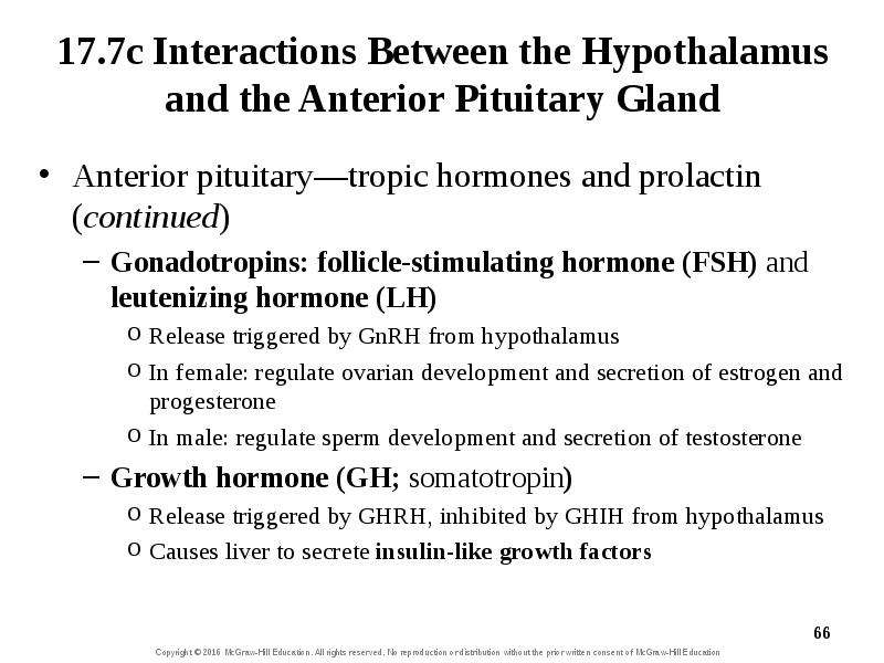
17.7c Interactions Between the Hypothalamus and the Anterior Pituitary Gland Anterior pituitary—tropic hormones and prolactin (continued) Gonadotropins: follicle-stimulating hormone (FSH) and leutenizing hormone (LH) Release triggered by GnRH from hypothalamus In female: regulate ovarian development and secretion of estrogen and progesterone In male: regulate sperm development and secretion of testosterone Growth hormone (GH; somatotropin) Release triggered by GHRH, inhibited by GHIH from hypothalamus Causes liver to secrete insulin-like growth factors

Слайд 67


chapt17_apr_lecture2e, слайд №67
Описание слайда:

Слайд 68


Clinical View: Hypophysectomy Surgical removal of the pituitary gland because of tumors Preferred surgical approach through nasal cavity Various...
Описание слайда:
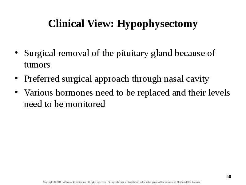
Clinical View: Hypophysectomy Surgical removal of the pituitary gland because of tumors Preferred surgical approach through nasal cavity Various hormones need to be replaced and their levels need to be monitored

Слайд 69


What did you learn? Where are secondary plexus blood vessels located? Where are tropic hormones synthesized and what is their general function? Where...
Описание слайда:
What did you learn? Where are secondary plexus blood vessels located? Where are tropic hormones synthesized and what is their general function? Where is oxytocin synthesized and where is it released?

Слайд 70


17.8 Representative Hormones Regulated by the Hypothalamus Describe the homeostatic system involving growth hormone. Describe thyroid gland location...
Описание слайда:
17.8 Representative Hormones Regulated by the Hypothalamus Describe the homeostatic system involving growth hormone. Describe thyroid gland location and anatomy. Discuss how thyroid hormones are produced, stored, and secreted. Explain the control of thyroid hormone by the hypothalamus and pituitary. Describe the structure and location of the adrenal glands.

Слайд 71


17.8 Representative Hormones Regulated by the Hypothalamus (continued) Name the three zones of the adrenal cortex and the hormones produced in each...
Описание слайда:
17.8 Representative Hormones Regulated by the Hypothalamus (continued) Name the three zones of the adrenal cortex and the hormones produced in each zone. Describe how the hypothalamus controls the release of glucocorticoid (cortisol) and the effects of cortisol.

Слайд 72


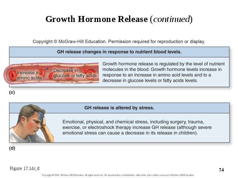
17.8a Growth Hormone Growth hormone (GH) functions include Stimulation of linear growth at epiphyseal plate Hypertrophy of muscle Release of...
Описание слайда:
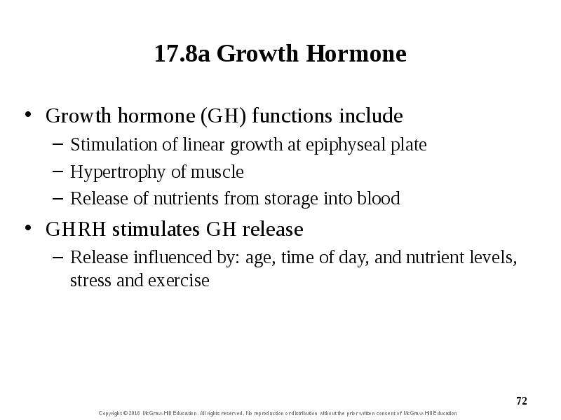
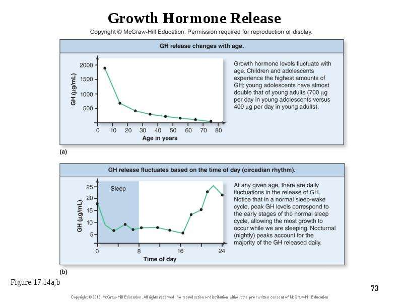
17.8a Growth Hormone Growth hormone (GH) functions include Stimulation of linear growth at epiphyseal plate Hypertrophy of muscle Release of nutrients from storage into blood GHRH stimulates GH release Release influenced by: age, time of day, and nutrient levels, stress and exercise

Слайд 73


Growth Hormone Release
Описание слайда:
Growth Hormone Release

Слайд 74


chapt17_apr_lecture2e, слайд №74
Описание слайда:

Слайд 75


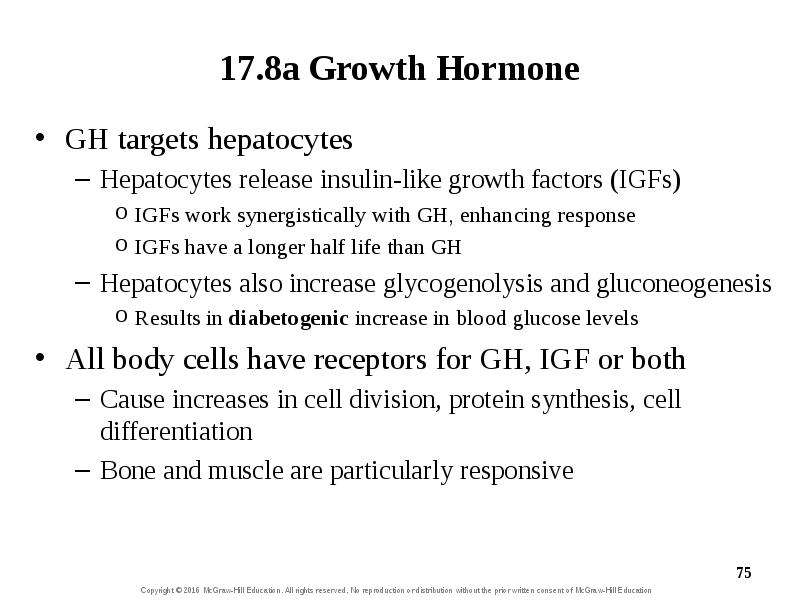
17.8a Growth Hormone GH targets hepatocytes Hepatocytes release insulin-like growth factors (IGFs) IGFs work synergistically with GH, enhancing...
Описание слайда:
17.8a Growth Hormone GH targets hepatocytes Hepatocytes release insulin-like growth factors (IGFs) IGFs work synergistically with GH, enhancing response IGFs have a longer half life than GH Hepatocytes also increase glycogenolysis and gluconeogenesis Results in diabetogenic increase in blood glucose levels All body cells have receptors for GH, IGF or both Cause increases in cell division, protein synthesis, cell differentiation Bone and muscle are particularly responsive

Слайд 76


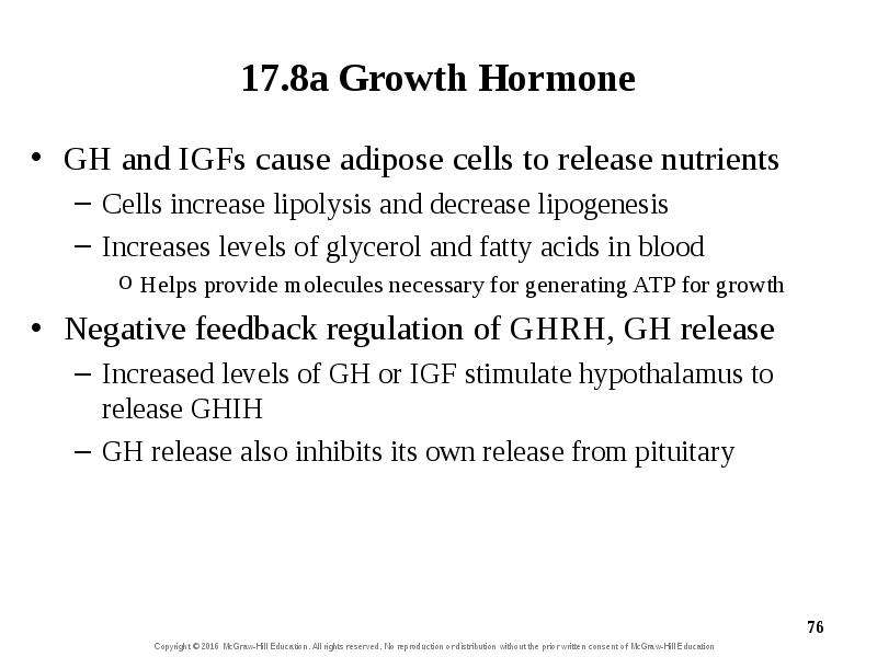
17.8a Growth Hormone GH and IGFs cause adipose cells to release nutrients Cells increase lipolysis and decrease lipogenesis Increases levels of...
Описание слайда:
17.8a Growth Hormone GH and IGFs cause adipose cells to release nutrients Cells increase lipolysis and decrease lipogenesis Increases levels of glycerol and fatty acids in blood Helps provide molecules necessary for generating ATP for growth Negative feedback regulation of GHRH, GH release Increased levels of GH or IGF stimulate hypothalamus to release GHIH GH release also inhibits its own release from pituitary

Слайд 77


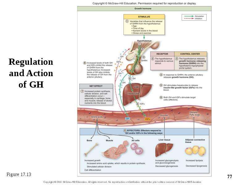
Regulation and Action of GH
Описание слайда:
Regulation and Action of GH

Слайд 78


Clinical View: Disorders of Growth Hormone Secretion Growth hormone deficiency (pituitary dwarfism) Inadequate growth hormone production Due to...
Описание слайда:
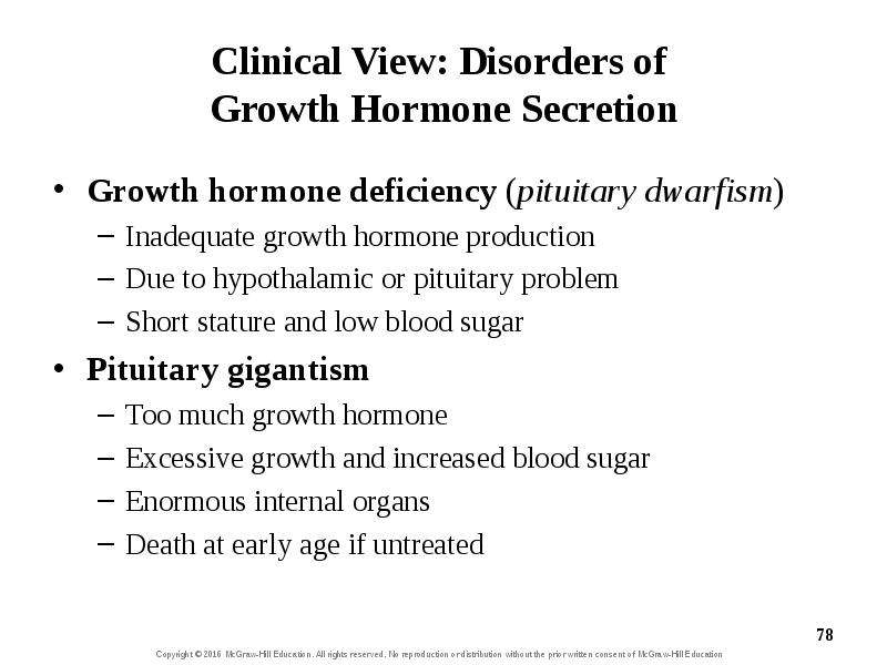
Clinical View: Disorders of Growth Hormone Secretion Growth hormone deficiency (pituitary dwarfism) Inadequate growth hormone production Due to hypothalamic or pituitary problem Short stature and low blood sugar Pituitary gigantism Too much growth hormone Excessive growth and increased blood sugar Enormous internal organs Death at early age if untreated

Слайд 79


Clinical View: Disorders of Growth Hormone Secretion (continued) Acromegaly Excessive growth hormone production in adult Enlargement of bones of...
Описание слайда:
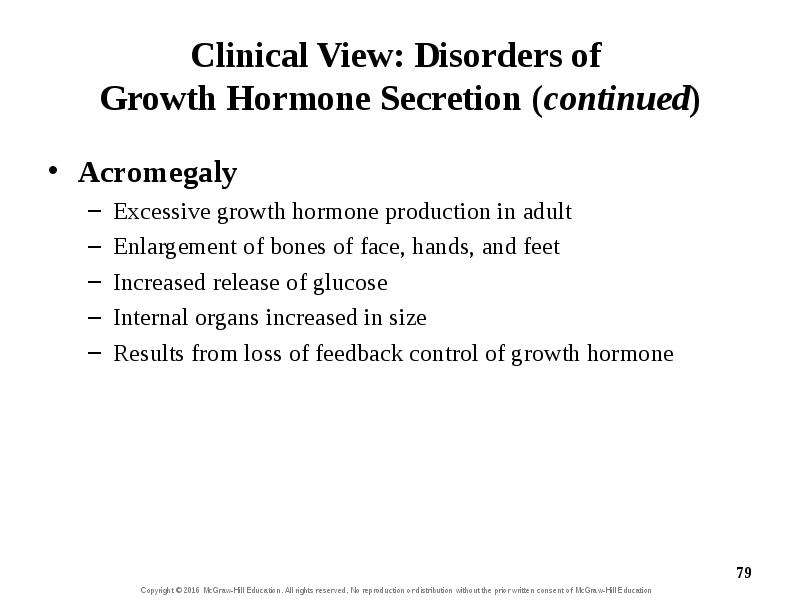
Clinical View: Disorders of Growth Hormone Secretion (continued) Acromegaly Excessive growth hormone production in adult Enlargement of bones of face, hands, and feet Increased release of glucose Internal organs increased in size Results from loss of feedback control of growth hormone

Слайд 80


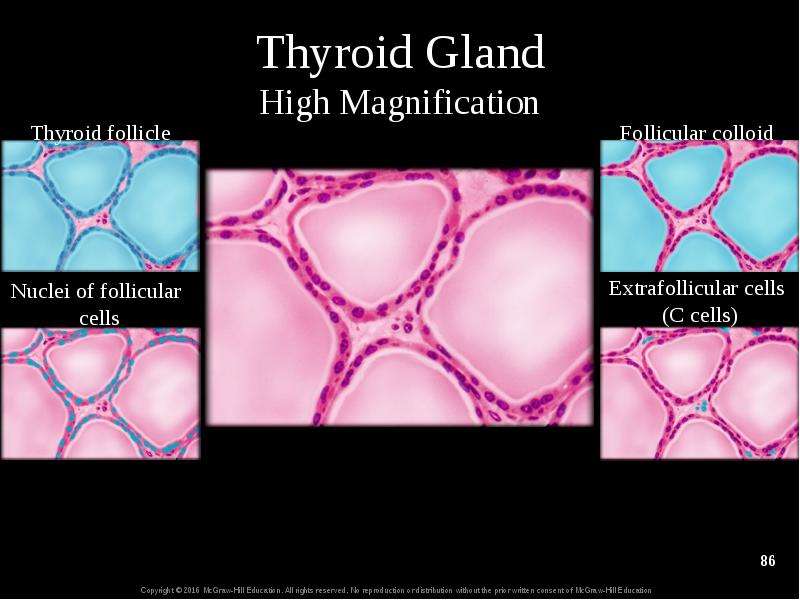
17.8b Thyroid Gland and Thyroid Hormone Anatomy of the thyroid gland Sits inferior to thyroid cartilage of larynx, anterior to trachea Left and right...
Описание слайда:
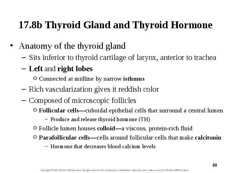
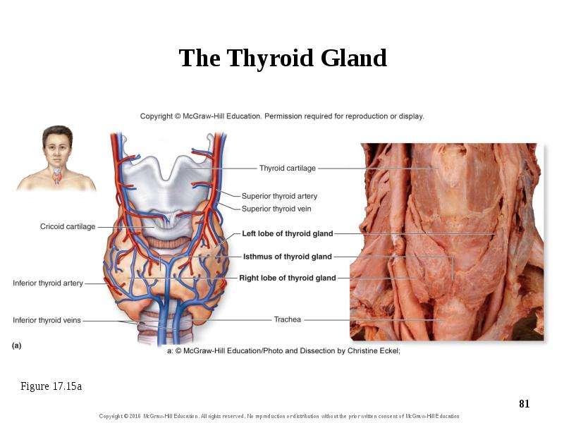
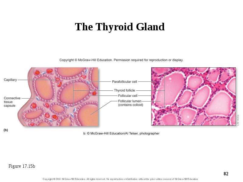
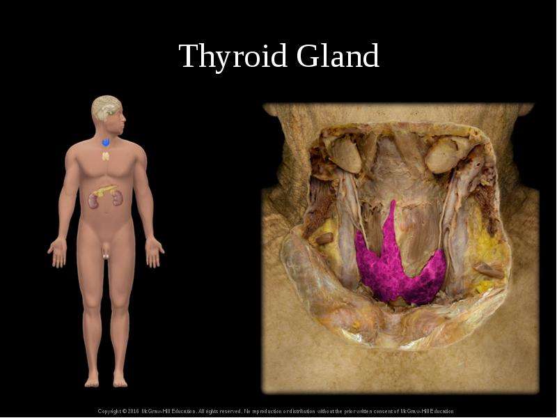
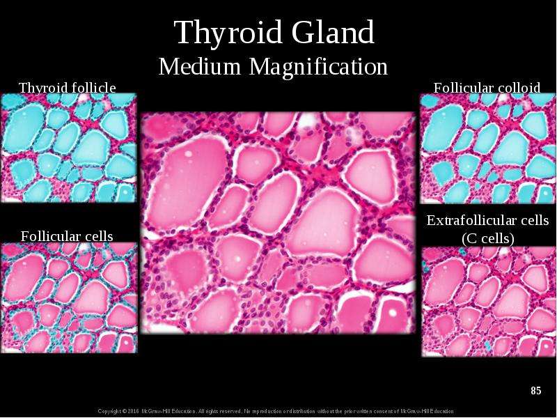
17.8b Thyroid Gland and Thyroid Hormone Anatomy of the thyroid gland Sits inferior to thyroid cartilage of larynx, anterior to trachea Left and right lobes Connected at midline by narrow isthmus Rich vascularization gives it reddish color Composed of microscopic follicles Follicular cells—cuboidal epithelial cells that surround a central lumen Produce and release thyroid hormone (TH) Follicle lumen houses colloid—a viscous, protein-rich fluid Parafollicular cells—cells around follicular cells that make calcitonin Hormone that decreases blood calcium levels

Слайд 81


The Thyroid Gland
Описание слайда:
The Thyroid Gland

Слайд 82


The Thyroid Gland
Описание слайда:
The Thyroid Gland

Слайд 83


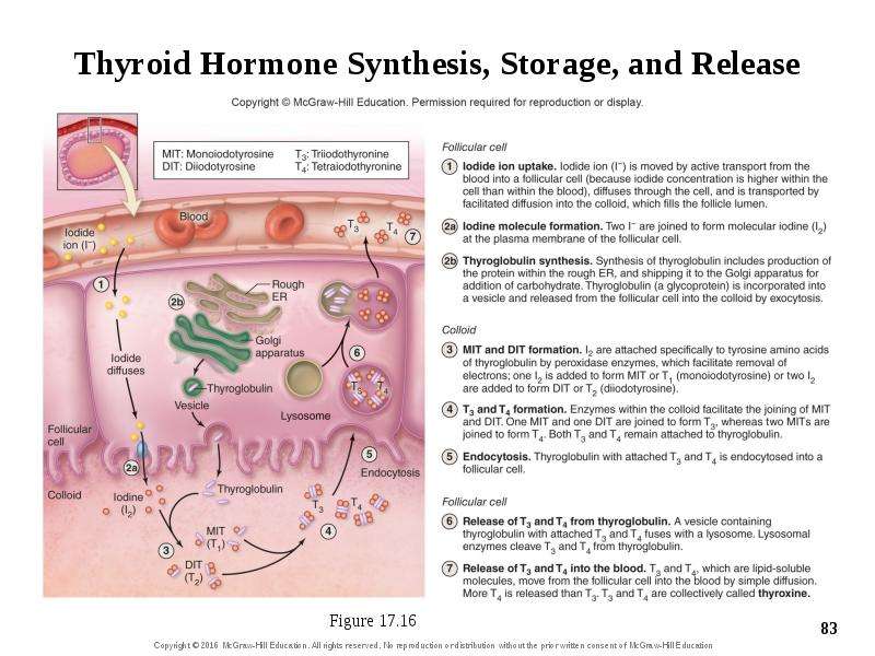
Thyroid Hormone Synthesis, Storage, and Release
Описание слайда:
Thyroid Hormone Synthesis, Storage, and Release

Слайд 84


Thyroid Gland
Описание слайда:
Thyroid Gland

Слайд 85


Thyroid Gland Medium Magnification
Описание слайда:
Thyroid Gland Medium Magnification

Слайд 86


Thyroid Gland High Magnification
Описание слайда:
Thyroid Gland High Magnification

Слайд 87


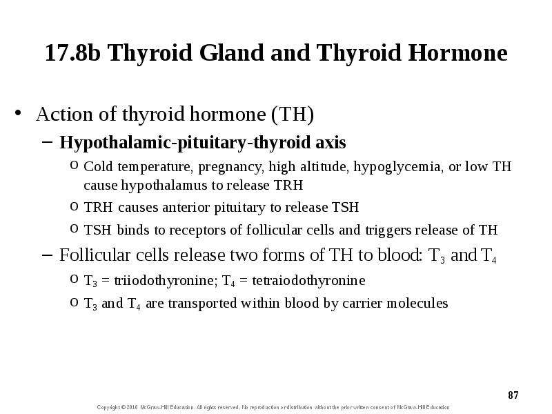
17.8b Thyroid Gland and Thyroid Hormone Action of thyroid hormone (TH) Hypothalamic-pituitary-thyroid axis Cold temperature, pregnancy, high...
Описание слайда:
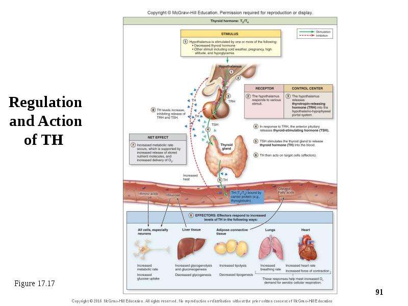
17.8b Thyroid Gland and Thyroid Hormone Action of thyroid hormone (TH) Hypothalamic-pituitary-thyroid axis Cold temperature, pregnancy, high altitude, hypoglycemia, or low TH cause hypothalamus to release TRH TRH causes anterior pituitary to release TSH TSH binds to receptors of follicular cells and triggers release of TH Follicular cells release two forms of TH to blood: T3 and T4 T3 = triiodothyronine; T4 = tetraiodothyronine T3 and T4 are transported within blood by carrier molecules

Слайд 88


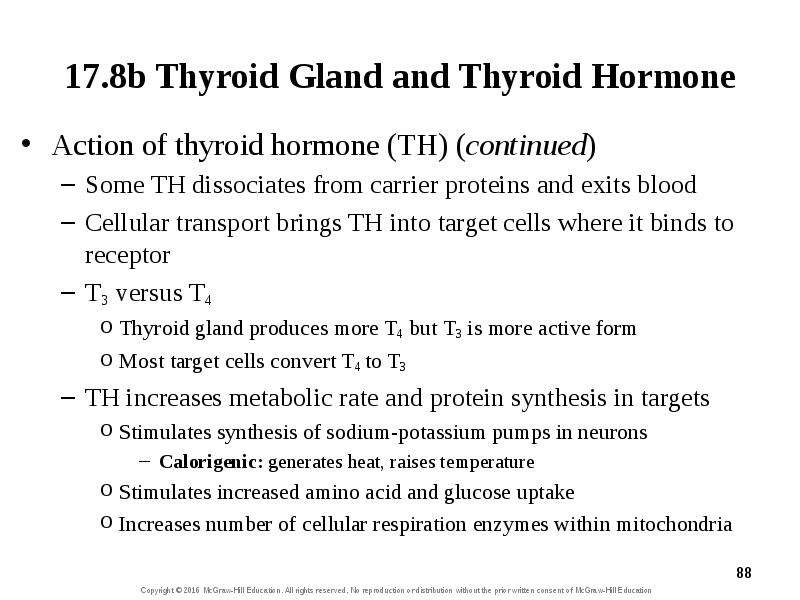
17.8b Thyroid Gland and Thyroid Hormone Action of thyroid hormone (TH) (continued) Some TH dissociates from carrier proteins and exits blood Cellular...
Описание слайда:
17.8b Thyroid Gland and Thyroid Hormone Action of thyroid hormone (TH) (continued) Some TH dissociates from carrier proteins and exits blood Cellular transport brings TH into target cells where it binds to receptor T3 versus T4 Thyroid gland produces more T4 but T3 is more active form Most target cells convert T4 to T3 TH increases metabolic rate and protein synthesis in targets Stimulates synthesis of sodium-potassium pumps in neurons Calorigenic: generates heat, raises temperature Stimulates increased amino acid and glucose uptake Increases number of cellular respiration enzymes within mitochondria

Слайд 89


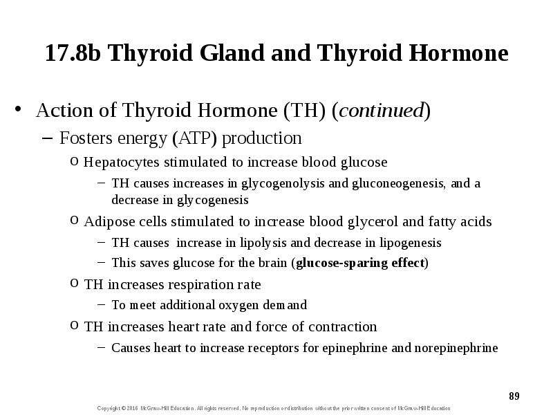
17.8b Thyroid Gland and Thyroid Hormone Action of Thyroid Hormone (TH) (continued) Fosters energy (ATP) production Hepatocytes stimulated to increase...
Описание слайда:
17.8b Thyroid Gland and Thyroid Hormone Action of Thyroid Hormone (TH) (continued) Fosters energy (ATP) production Hepatocytes stimulated to increase blood glucose TH causes increases in glycogenolysis and gluconeogenesis, and a decrease in glycogenesis Adipose cells stimulated to increase blood glycerol and fatty acids TH causes increase in lipolysis and decrease in lipogenesis This saves glucose for the brain (glucose-sparing effect) TH increases respiration rate To meet additional oxygen demand TH increases heart rate and force of contraction Causes heart to increase receptors for epinephrine and norepinephrine

Слайд 90


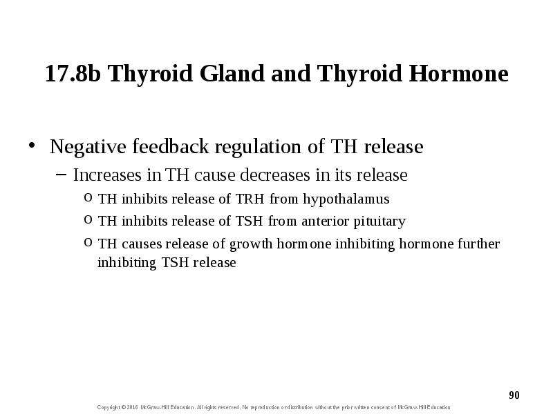
17.8b Thyroid Gland and Thyroid Hormone Negative feedback regulation of TH release Increases in TH cause decreases in its release TH inhibits release...
Описание слайда:
17.8b Thyroid Gland and Thyroid Hormone Negative feedback regulation of TH release Increases in TH cause decreases in its release TH inhibits release of TRH from hypothalamus TH inhibits release of TSH from anterior pituitary TH causes release of growth hormone inhibiting hormone further inhibiting TSH release

Слайд 91


Regulation and Action of TH
Описание слайда:
Regulation and Action of TH

Слайд 92


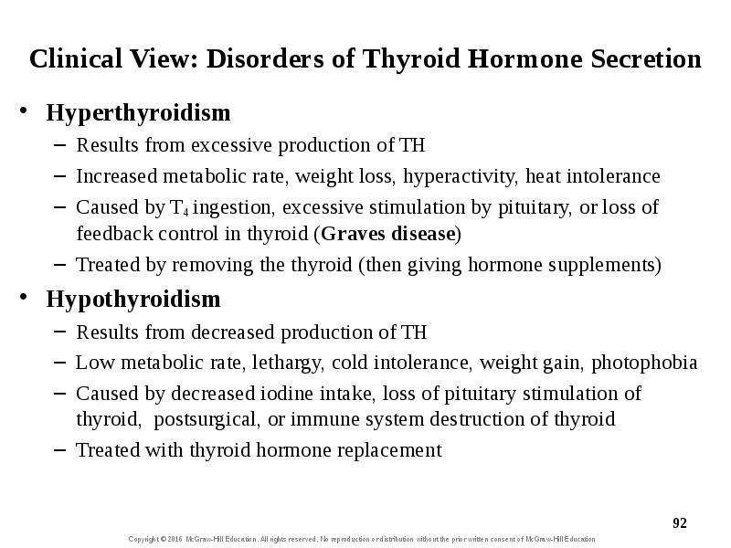
Clinical View: Disorders of Thyroid Hormone Secretion Hyperthyroidism Results from excessive production of TH Increased metabolic rate, weight loss,...
Описание слайда:
Clinical View: Disorders of Thyroid Hormone Secretion Hyperthyroidism Results from excessive production of TH Increased metabolic rate, weight loss, hyperactivity, heat intolerance Caused by T4 ingestion, excessive stimulation by pituitary, or loss of feedback control in thyroid (Graves disease) Treated by removing the thyroid (then giving hormone supplements) Hypothyroidism Results from decreased production of TH Low metabolic rate, lethargy, cold intolerance, weight gain, photophobia Caused by decreased iodine intake, loss of pituitary stimulation of thyroid, postsurgical, or immune system destruction of thyroid Treated with thyroid hormone replacement

Слайд 93


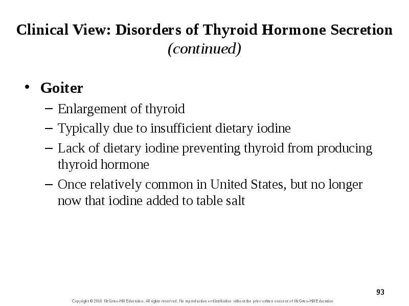
Clinical View: Disorders of Thyroid Hormone Secretion (continued) Goiter Enlargement of thyroid Typically due to insufficient dietary iodine Lack of...
Описание слайда:
Clinical View: Disorders of Thyroid Hormone Secretion (continued) Goiter Enlargement of thyroid Typically due to insufficient dietary iodine Lack of dietary iodine preventing thyroid from producing thyroid hormone Once relatively common in United States, but no longer now that iodine added to table salt

Слайд 94


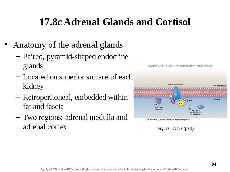
17.8c Adrenal Glands and Cortisol Anatomy of the adrenal glands Paired, pyramid-shaped endocrine glands Located on superior surface of each kidney...
Описание слайда:
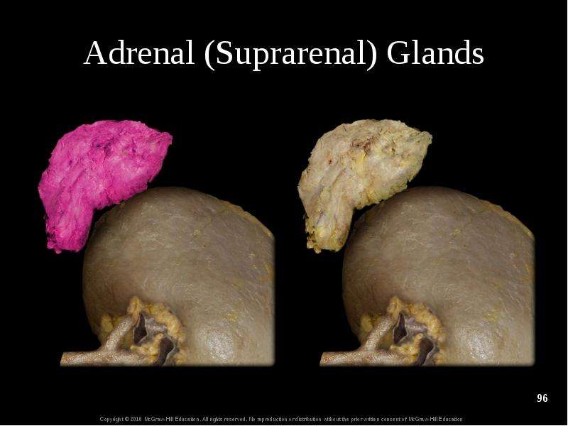
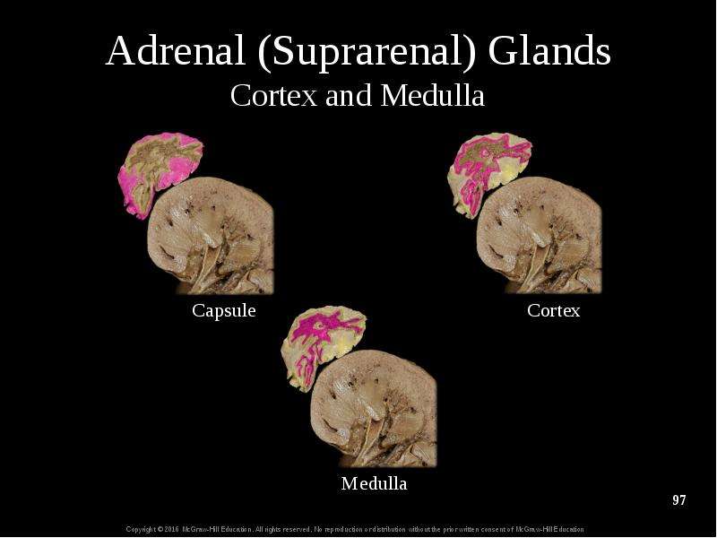
17.8c Adrenal Glands and Cortisol Anatomy of the adrenal glands Paired, pyramid-shaped endocrine glands Located on superior surface of each kidney Retroperitoneal, embedded within fat and fascia Two regions: adrenal medulla and adrenal cortex

Слайд 95


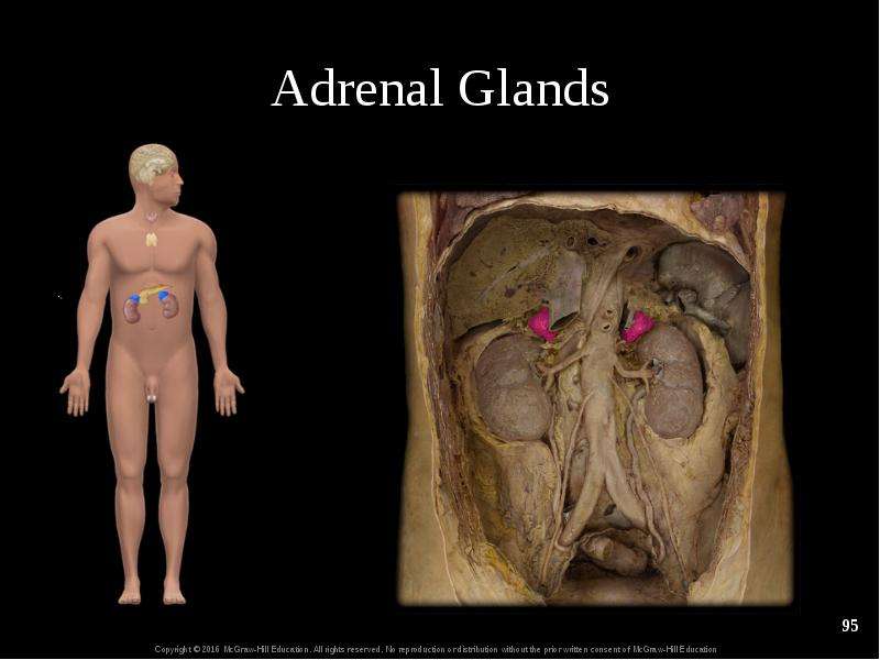
Adrenal Glands
Описание слайда:
Adrenal Glands

Слайд 96


Adrenal (Suprarenal) Glands
Описание слайда:
Adrenal (Suprarenal) Glands

Слайд 97


Adrenal (Suprarenal) Glands Cortex and Medulla
Описание слайда:
Adrenal (Suprarenal) Glands Cortex and Medulla

Слайд 98


17.8c Adrenal Glands and Cortisol Anatomy of the adrenal glands (continued) Adrenal medulla Forms inner core of each adrenal gland Red-brown color...
Описание слайда:
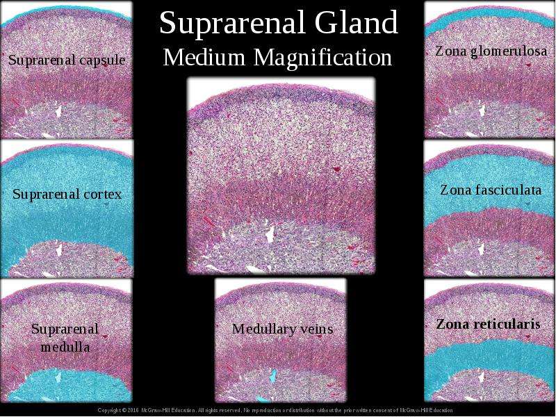
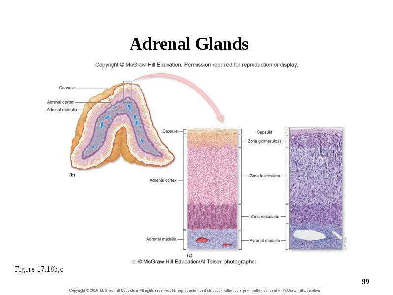
17.8c Adrenal Glands and Cortisol Anatomy of the adrenal glands (continued) Adrenal medulla Forms inner core of each adrenal gland Red-brown color due to extensive blood vessels Releases epinephrine and norepinephrine with sympathetic stimulation Adrenal cortex Synthesizes more than 25 corticosteroids Yellow color due to lipids within cells Three regions producing different steroid hormones: zona glomerulosa, zona fasciculata, and the inner zona reticularis

Слайд 99


Adrenal Glands
Описание слайда:
Adrenal Glands

Слайд 100


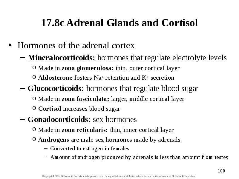
17.8c Adrenal Glands and Cortisol Hormones of the adrenal cortex Mineralocorticoids: hormones that regulate electrolyte levels Made in zona...
Описание слайда:
17.8c Adrenal Glands and Cortisol Hormones of the adrenal cortex Mineralocorticoids: hormones that regulate electrolyte levels Made in zona glomerulosa: thin, outer cortical layer Aldosterone fosters Na+ retention and K+ secretion Glucocorticoids: hormones that regulate blood sugar Made in zona fasciculata: larger, middle cortical layer Cortisol increases blood sugar Gonadocorticoids: sex hormones Made in zona reticularis: thin, inner cortical layer Androgens are male sex hormones made by adrenals Converted to estrogen in females Amount of androgen produced by adrenals is less than amount from testes

Слайд 101


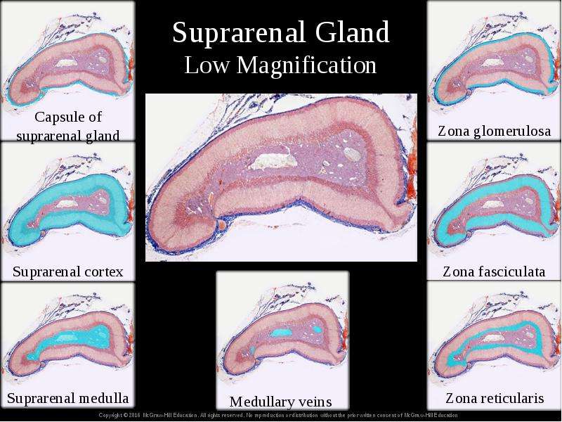
Suprarenal Gland Low Magnification
Описание слайда:
Suprarenal Gland Low Magnification

Слайд 102


Suprarenal Gland Medium Magnification
Описание слайда:
Suprarenal Gland Medium Magnification

Слайд 103


17.8c Adrenal Glands and Cortisol Action of cortisol Cortisol and corticosterone increase nutrient levels in blood To resist stress and repair...
Описание слайда:
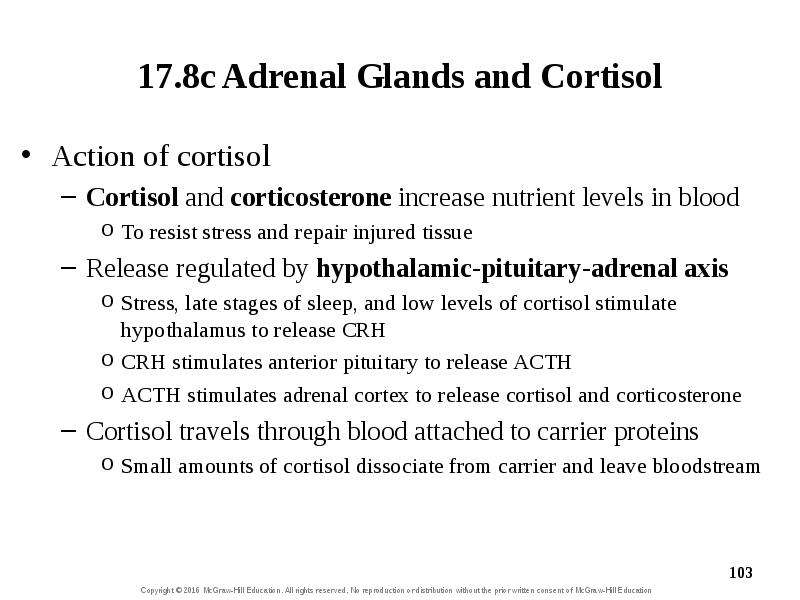
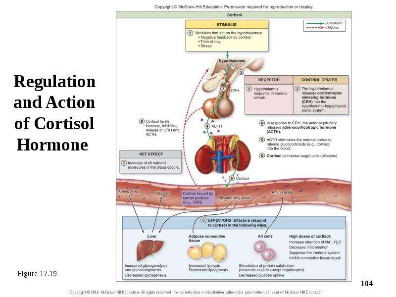
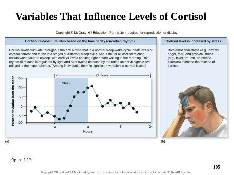
17.8c Adrenal Glands and Cortisol Action of cortisol Cortisol and corticosterone increase nutrient levels in blood To resist stress and repair injured tissue Release regulated by hypothalamic-pituitary-adrenal axis Stress, late stages of sleep, and low levels of cortisol stimulate hypothalamus to release CRH CRH stimulates anterior pituitary to release ACTH ACTH stimulates adrenal cortex to release cortisol and corticosterone Cortisol travels through blood attached to carrier proteins Small amounts of cortisol dissociate from carrier and leave bloodstream

Слайд 104


Regulation and Action of Cortisol Hormone
Описание слайда:
Regulation and Action of Cortisol Hormone

Слайд 105


Variables That Influence Levels of Cortisol
Описание слайда:
Variables That Influence Levels of Cortisol

Слайд 106


17.8c Adrenal Glands and Cortisol Action of cortisol (continued) Cortisol diffuses through target cell’s membrane and binds to intracellular receptor...
Описание слайда:
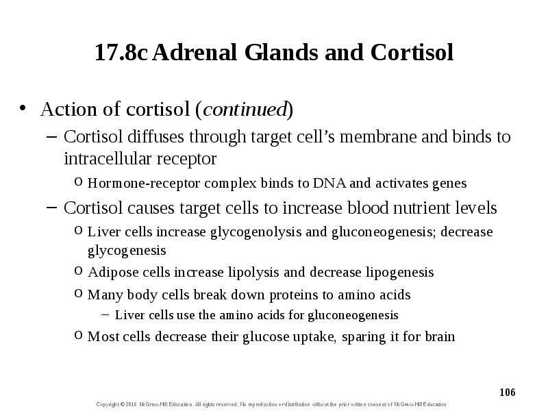
17.8c Adrenal Glands and Cortisol Action of cortisol (continued) Cortisol diffuses through target cell’s membrane and binds to intracellular receptor Hormone-receptor complex binds to DNA and activates genes Cortisol causes target cells to increase blood nutrient levels Liver cells increase glycogenolysis and gluconeogenesis; decrease glycogenesis Adipose cells increase lipolysis and decrease lipogenesis Many body cells break down proteins to amino acids Liver cells use the amino acids for gluconeogenesis Most cells decrease their glucose uptake, sparing it for brain

Слайд 107


17.8c Adrenal Glands and Cortisol Cortisol levels are regulated by negative feedback Cortisol inhibits release of CRH from hypothalamus and ACTH from...
Описание слайда:
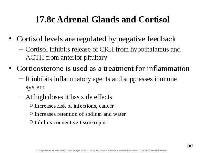
17.8c Adrenal Glands and Cortisol Cortisol levels are regulated by negative feedback Cortisol inhibits release of CRH from hypothalamus and ACTH from anterior pituitary Corticosterone is used as a treatment for inflammation It inhibits inflammatory agents and suppresses immune system At high doses it has side effects Increases risk of infections, cancer Increases retention of sodium and water Inhibits connective tissue repair

Слайд 108


Clinical View: Disorders in Adrenal Cortex Hormone Secretion Cushing syndrome Chronic exposure to excessive glucocorticoid hormones in people taking...
Описание слайда:
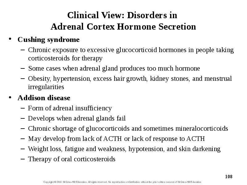
Clinical View: Disorders in Adrenal Cortex Hormone Secretion Cushing syndrome Chronic exposure to excessive glucocorticoid hormones in people taking corticosteroids for therapy Some cases when adrenal gland produces too much hormone Obesity, hypertension, excess hair growth, kidney stones, and menstrual irregularities Addison disease Form of adrenal insufficiency Develops when adrenal glands fail Chronic shortage of glucocorticoids and sometimes mineralocorticoids May develop from lack of ACTH or lack of response to ACTH Weight loss, fatigue and weakness, hypotension, and skin darkening Therapy of oral corticosteroids

Слайд 109


Clinical View: Disorders in Adrenal Cortex Hormone Secretion (continued) Adrenogenital syndrome (congenital adrenal hyperplasia) Begins in embryo or...
Описание слайда:
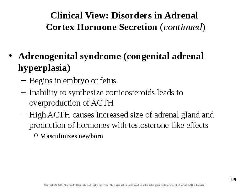
Clinical View: Disorders in Adrenal Cortex Hormone Secretion (continued) Adrenogenital syndrome (congenital adrenal hyperplasia) Begins in embryo or fetus Inability to synthesize corticosteroids leads to overproduction of ACTH High ACTH causes increased size of adrenal gland and production of hormones with testosterone-like effects Masculinizes newborn

Слайд 110


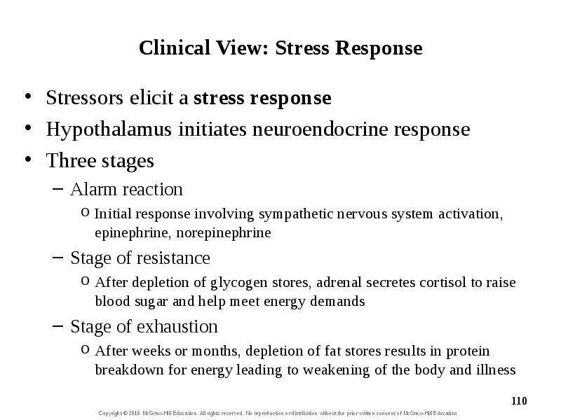
Clinical View: Stress Response Stressors elicit a stress response Hypothalamus initiates neuroendocrine response Three stages Alarm reaction Initial...
Описание слайда:
Clinical View: Stress Response Stressors elicit a stress response Hypothalamus initiates neuroendocrine response Three stages Alarm reaction Initial response involving sympathetic nervous system activation, epinephrine, norepinephrine Stage of resistance After depletion of glycogen stores, adrenal secretes cortisol to raise blood sugar and help meet energy demands Stage of exhaustion After weeks or months, depletion of fat stores results in protein breakdown for energy leading to weakening of the body and illness

Слайд 111


What did you learn? At what time of day are growth hormone levels highest? What is the function of thyroid follicular cells? What is the primary...
Описание слайда:
What did you learn? At what time of day are growth hormone levels highest? What is the function of thyroid follicular cells? What is the primary mineralocorticoid and what are its specific effects?

Слайд 112


17.9 Pancreatic Hormones Describe the gross anatomy and cellular structure of the pancreas. Identify the primary types of pancreatic islet cells and...
Описание слайда:
17.9 Pancreatic Hormones Describe the gross anatomy and cellular structure of the pancreas. Identify the primary types of pancreatic islet cells and the hormones they produce. Describe the action of insulin in lowering blood glucose concentration. Explain the action of glucagon in raising blood glucose concentration.

Слайд 113


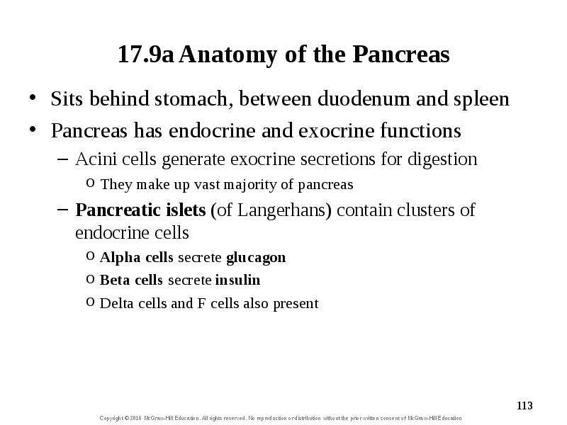
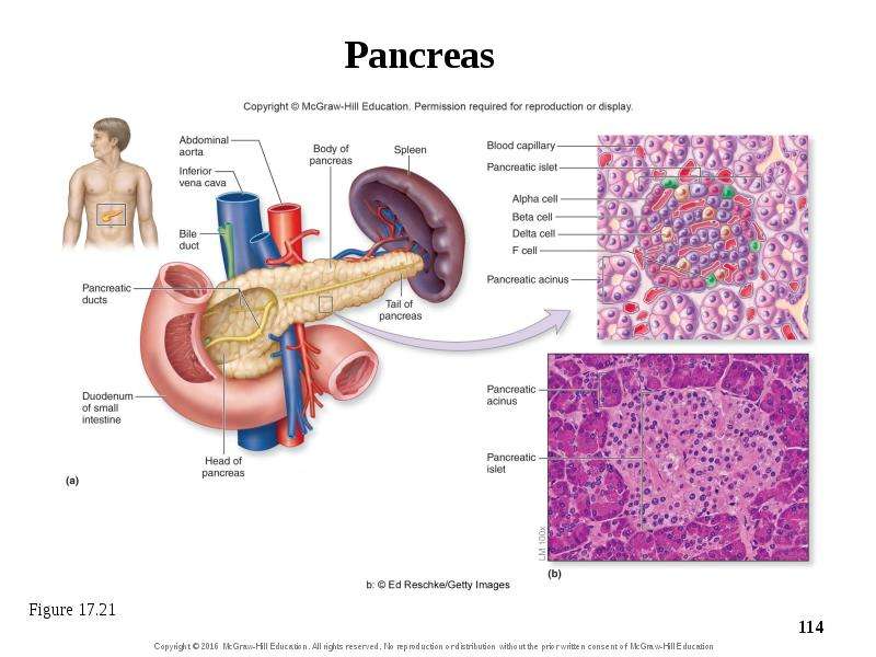
17.9a Anatomy of the Pancreas Sits behind stomach, between duodenum and spleen Pancreas has endocrine and exocrine functions Acini cells generate...
Описание слайда:
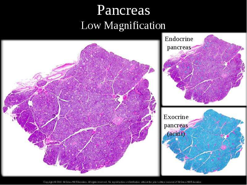
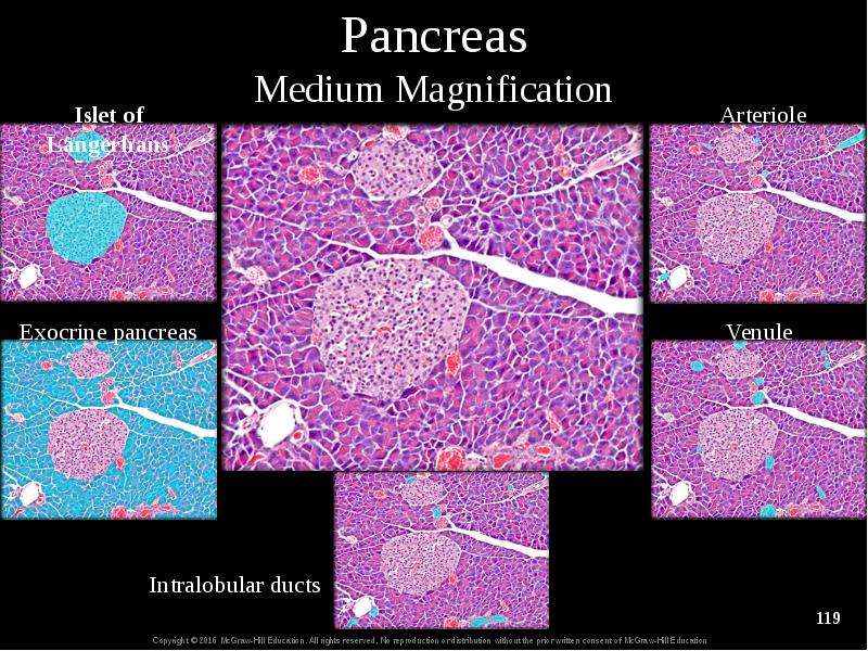
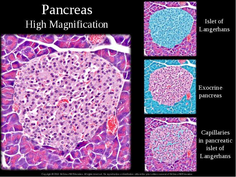
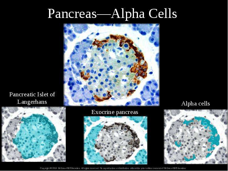
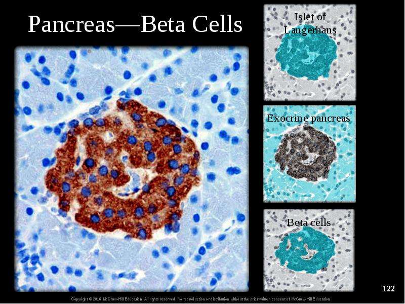
17.9a Anatomy of the Pancreas Sits behind stomach, between duodenum and spleen Pancreas has endocrine and exocrine functions Acini cells generate exocrine secretions for digestion They make up vast majority of pancreas Pancreatic islets (of Langerhans) contain clusters of endocrine cells Alpha cells secrete glucagon Beta cells secrete insulin Delta cells and F cells also present

Слайд 114


Pancreas
Описание слайда:
Pancreas

Слайд 115


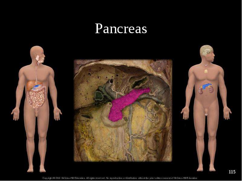
Pancreas
Описание слайда:
Pancreas

Слайд 116


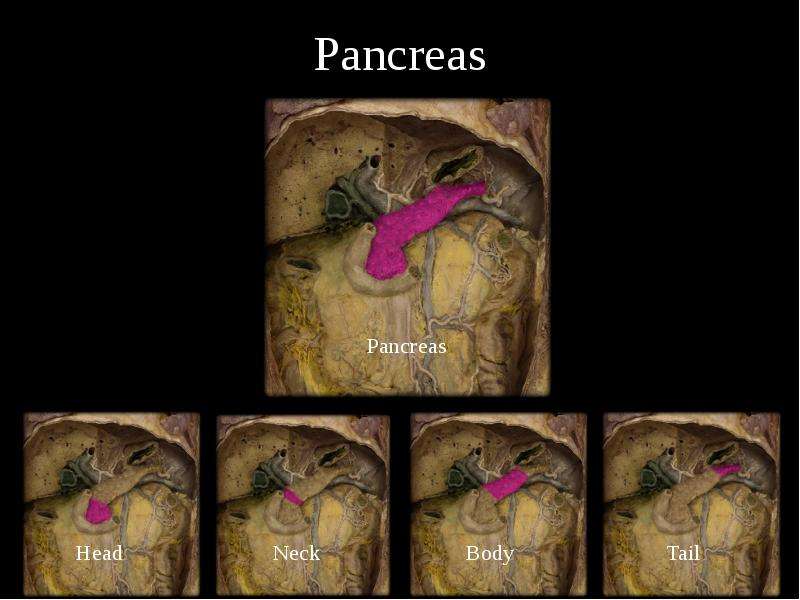
Pancreas
Описание слайда:
Pancreas

Слайд 117


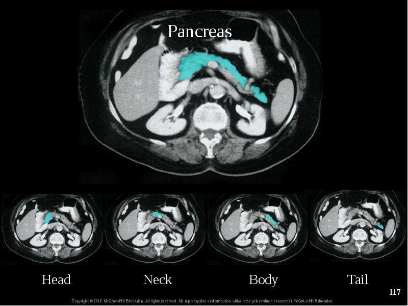
chapt17_apr_lecture2e, слайд №117
Описание слайда:

Слайд 118


Pancreas Low Magnification
Описание слайда:
Pancreas Low Magnification

Слайд 119


Pancreas Medium Magnification
Описание слайда:
Pancreas Medium Magnification

Слайд 120


Pancreas High Magnification
Описание слайда:
Pancreas High Magnification

Слайд 121


Pancreas—Alpha Cells
Описание слайда:
Pancreas—Alpha Cells

Слайд 122


Pancreas—Beta Cells
Описание слайда:
Pancreas—Beta Cells

Слайд 123


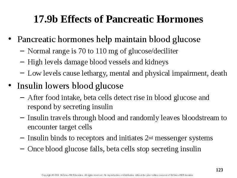
17.9b Effects of Pancreatic Hormones Pancreatic hormones help maintain blood glucose Normal range is 70 to 110 mg of glucose/deciliter High levels...
Описание слайда:
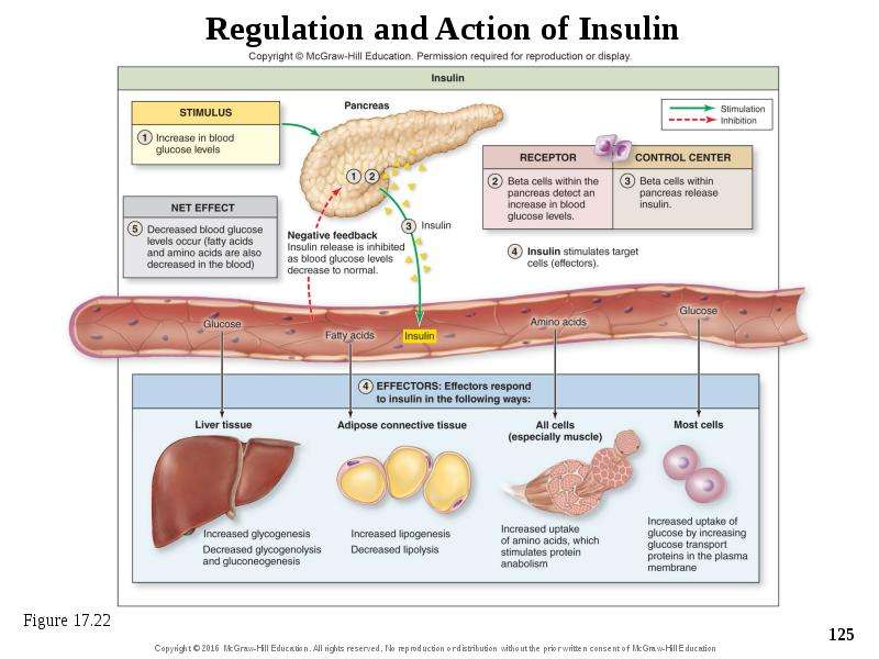
17.9b Effects of Pancreatic Hormones Pancreatic hormones help maintain blood glucose Normal range is 70 to 110 mg of glucose/deciliter High levels damage blood vessels and kidneys Low levels cause lethargy, mental and physical impairment, death Insulin lowers blood glucose After food intake, beta cells detect rise in blood glucose and respond by secreting insulin Insulin travels through blood and randomly leaves bloodstream to encounter target cells Insulin binds to receptors and initiates 2nd messenger systems Once blood glucose falls, beta cells stop secreting insulin

Слайд 124


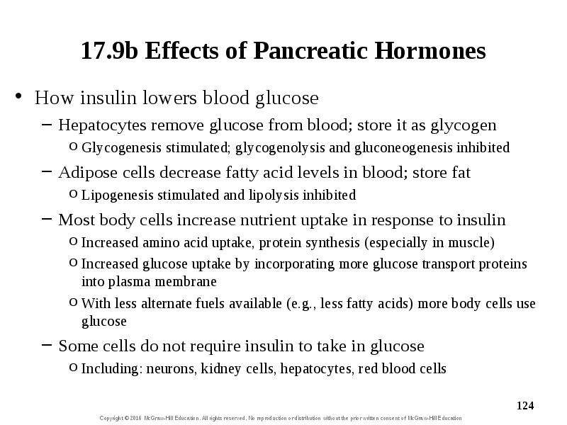
17.9b Effects of Pancreatic Hormones How insulin lowers blood glucose Hepatocytes remove glucose from blood; store it as glycogen Glycogenesis...
Описание слайда:
17.9b Effects of Pancreatic Hormones How insulin lowers blood glucose Hepatocytes remove glucose from blood; store it as glycogen Glycogenesis stimulated; glycogenolysis and gluconeogenesis inhibited Adipose cells decrease fatty acid levels in blood; store fat Lipogenesis stimulated and lipolysis inhibited Most body cells increase nutrient uptake in response to insulin Increased amino acid uptake, protein synthesis (especially in muscle) Increased glucose uptake by incorporating more glucose transport proteins into plasma membrane With less alternate fuels available (e.g., less fatty acids) more body cells use glucose Some cells do not require insulin to take in glucose Including: neurons, kidney cells, hepatocytes, red blood cells

Слайд 125


Regulation and Action of Insulin
Описание слайда:
Regulation and Action of Insulin

Слайд 126


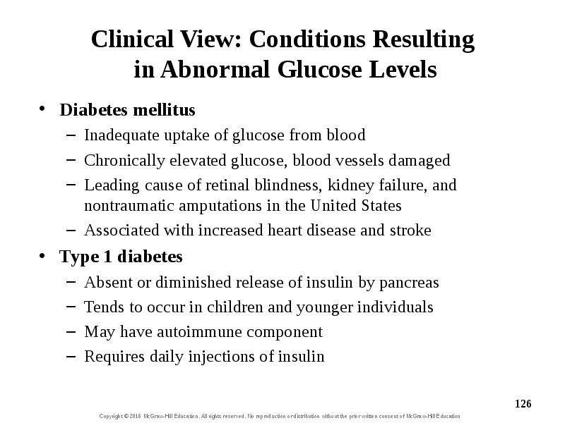
Clinical View: Conditions Resulting in Abnormal Glucose Levels Diabetes mellitus Inadequate uptake of glucose from blood Chronically elevated...
Описание слайда:
Clinical View: Conditions Resulting in Abnormal Glucose Levels Diabetes mellitus Inadequate uptake of glucose from blood Chronically elevated glucose, blood vessels damaged Leading cause of retinal blindness, kidney failure, and nontraumatic amputations in the United States Associated with increased heart disease and stroke Type 1 diabetes Absent or diminished release of insulin by pancreas Tends to occur in children and younger individuals May have autoimmune component Requires daily injections of insulin

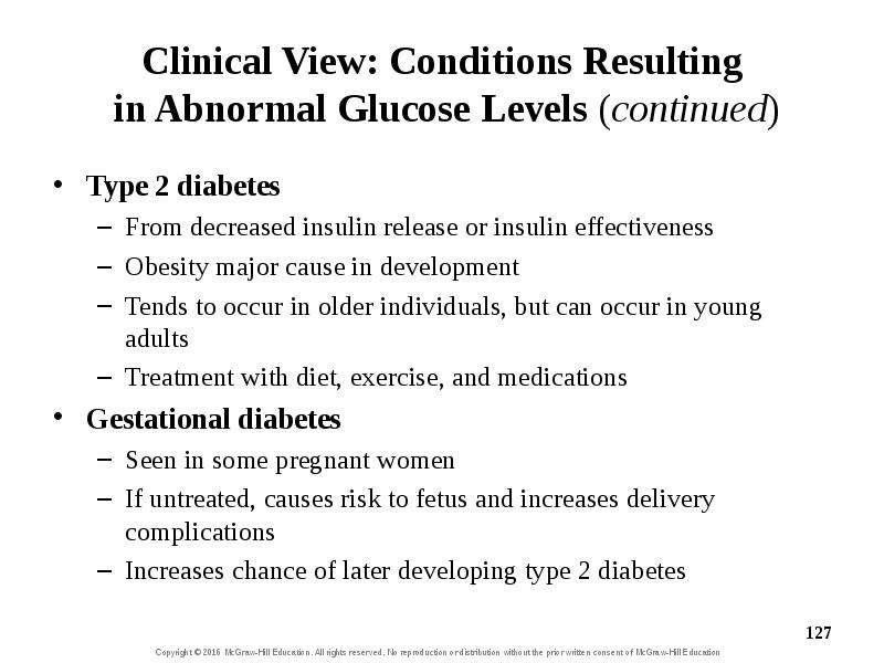
Слайд 127


Clinical View: Conditions Resulting in Abnormal Glucose Levels (continued) Type 2 diabetes From decreased insulin release or insulin effectiveness...
Описание слайда:
Clinical View: Conditions Resulting in Abnormal Glucose Levels (continued) Type 2 diabetes From decreased insulin release or insulin effectiveness Obesity major cause in development Tends to occur in older individuals, but can occur in young adults Treatment with diet, exercise, and medications Gestational diabetes Seen in some pregnant women If untreated, causes risk to fetus and increases delivery complications Increases chance of later developing type 2 diabetes

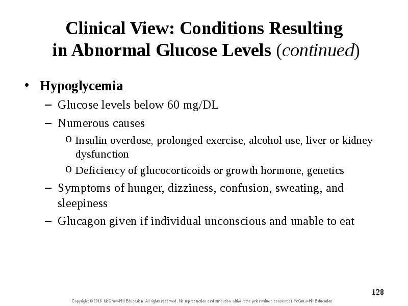
Слайд 128


Clinical View: Conditions Resulting in Abnormal Glucose Levels (continued) Hypoglycemia Glucose levels below 60 mg/DL Numerous causes Insulin...
Описание слайда:
Clinical View: Conditions Resulting in Abnormal Glucose Levels (continued) Hypoglycemia Glucose levels below 60 mg/DL Numerous causes Insulin overdose, prolonged exercise, alcohol use, liver or kidney dysfunction Deficiency of glucocorticoids or growth hormone, genetics Symptoms of hunger, dizziness, confusion, sweating, and sleepiness Glucagon given if individual unconscious and unable to eat

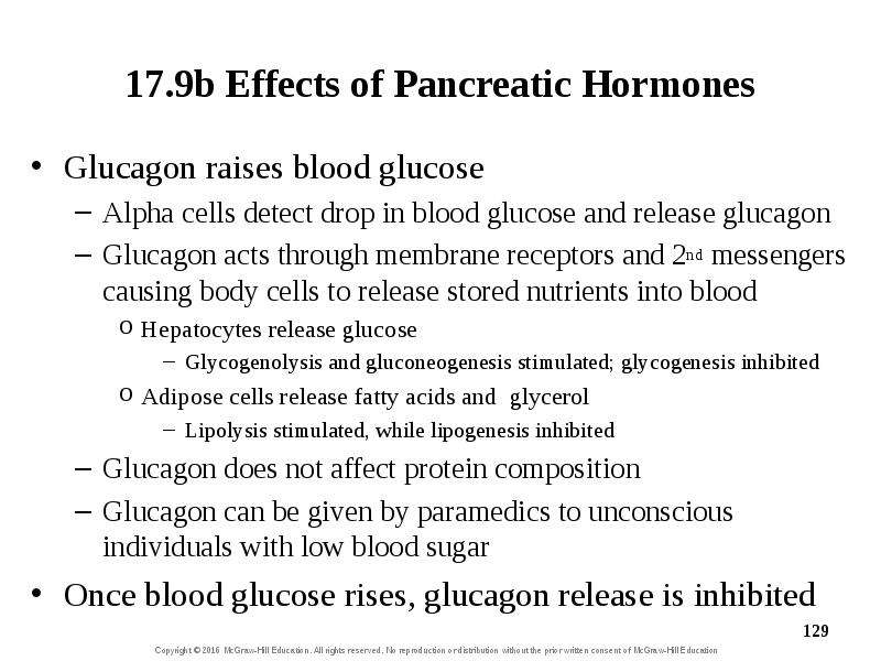
Слайд 129


17.9b Effects of Pancreatic Hormones Glucagon raises blood glucose Alpha cells detect drop in blood glucose and release glucagon Glucagon acts...
Описание слайда:
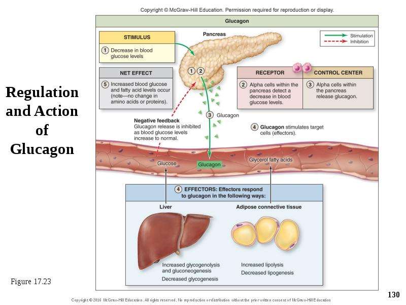
17.9b Effects of Pancreatic Hormones Glucagon raises blood glucose Alpha cells detect drop in blood glucose and release glucagon Glucagon acts through membrane receptors and 2nd messengers causing body cells to release stored nutrients into blood Hepatocytes release glucose Glycogenolysis and gluconeogenesis stimulated; glycogenesis inhibited Adipose cells release fatty acids and glycerol Lipolysis stimulated, while lipogenesis inhibited Glucagon does not affect protein composition Glucagon can be given by paramedics to unconscious individuals with low blood sugar Once blood glucose rises, glucagon release is inhibited

Слайд 130


Regulation and Action of Glucagon
Описание слайда:
Regulation and Action of Glucagon

Слайд 131


What did you learn? What function is served by the pancreatic islets? What effect would a decrease in insulin levels be expected to have on blood...
Описание слайда:
What did you learn? What function is served by the pancreatic islets? What effect would a decrease in insulin levels be expected to have on blood sugar? How is it that changes in the levels of fatty acids in the blood can affect blood sugar levels?

Слайд 132


17.10 Other Endocrine Glands Describe the general structure, location, and function of the pineal gland. Describe the general structure, location,...
Описание слайда:
17.10 Other Endocrine Glands Describe the general structure, location, and function of the pineal gland. Describe the general structure, location, and function of the parathyroid glands. Identify and provide a description of the general function of the hormone(s) released from each of the organs discussed in this section.

Слайд 133


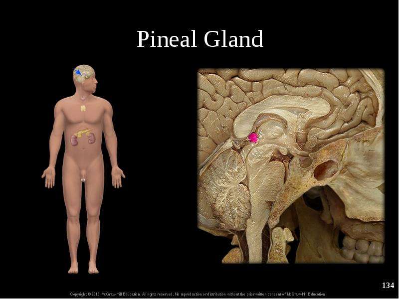
17.10a Pineal Gland Pineal gland is a small unpaired body in the epithalamus of the diencephalon Pineal secretes melatonin at night Causes drowsiness...
Описание слайда:
17.10a Pineal Gland Pineal gland is a small unpaired body in the epithalamus of the diencephalon Pineal secretes melatonin at night Causes drowsiness Regulates circadian rhythm and has effects on mood Melatonin influences GnRH secretion Has poorly understood effects on reproductive physiology

Слайд 134


Pineal Gland
Описание слайда:
Pineal Gland

Слайд 135


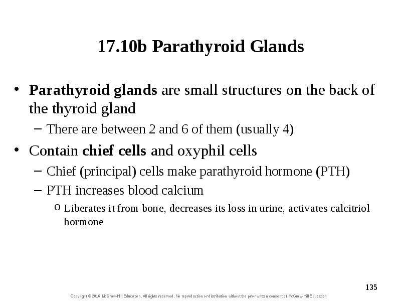
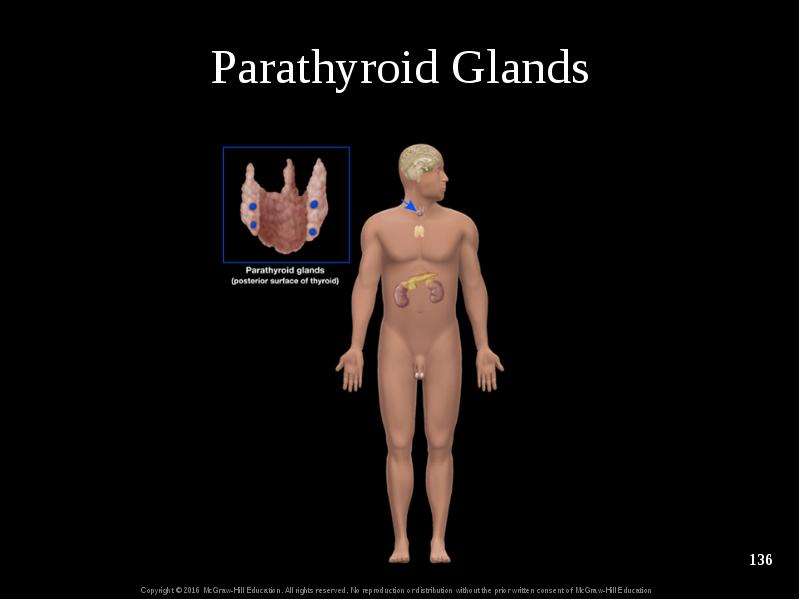
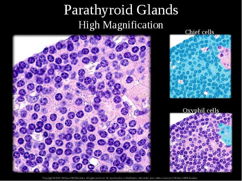
17.10b Parathyroid Glands Parathyroid glands are small structures on the back of the thyroid gland There are between 2 and 6 of them (usually 4)...
Описание слайда:
17.10b Parathyroid Glands Parathyroid glands are small structures on the back of the thyroid gland There are between 2 and 6 of them (usually 4) Contain chief cells and oxyphil cells Chief (principal) cells make parathyroid hormone (PTH) PTH increases blood calcium Liberates it from bone, decreases its loss in urine, activates calcitriol hormone

Слайд 136


Parathyroid Glands
Описание слайда:
Parathyroid Glands

Слайд 137


Parathyroid Glands High Magnification
Описание слайда:
Parathyroid Glands High Magnification

Слайд 138


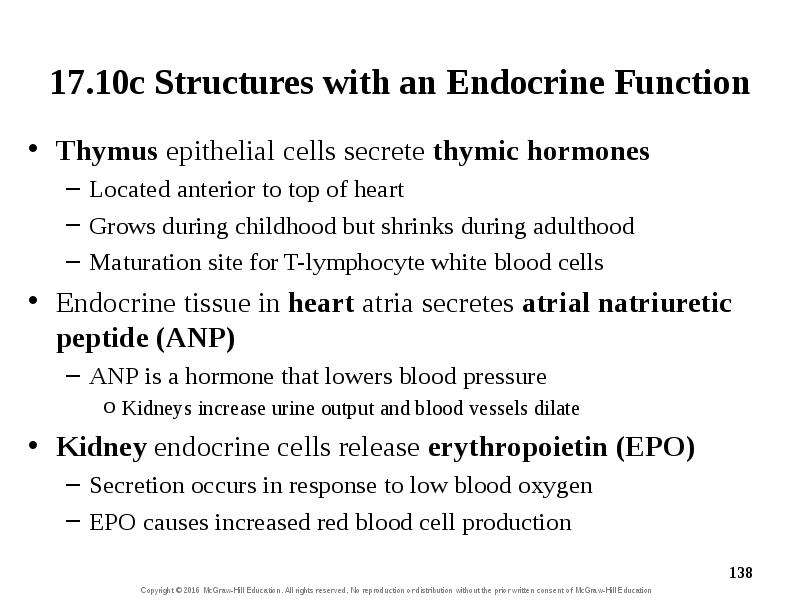
17.10c Structures with an Endocrine Function Thymus epithelial cells secrete thymic hormones Located anterior to top of heart Grows during childhood...
Описание слайда:
17.10c Structures with an Endocrine Function Thymus epithelial cells secrete thymic hormones Located anterior to top of heart Grows during childhood but shrinks during adulthood Maturation site for T-lymphocyte white blood cells Endocrine tissue in heart atria secretes atrial natriuretic peptide (ANP) ANP is a hormone that lowers blood pressure Kidneys increase urine output and blood vessels dilate Kidney endocrine cells release erythropoietin (EPO) Secretion occurs in response to low blood oxygen EPO causes increased red blood cell production

Слайд 139


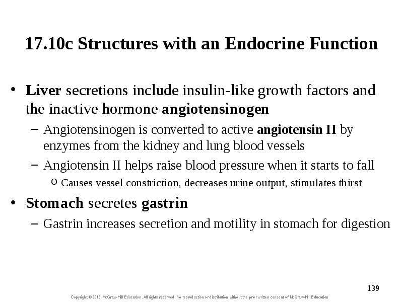
17.10c Structures with an Endocrine Function Liver secretions include insulin-like growth factors and the inactive hormone angiotensinogen...
Описание слайда:
17.10c Structures with an Endocrine Function Liver secretions include insulin-like growth factors and the inactive hormone angiotensinogen Angiotensinogen is converted to active angiotensin II by enzymes from the kidney and lung blood vessels Angiotensin II helps raise blood pressure when it starts to fall Causes vessel constriction, decreases urine output, stimulates thirst Stomach secretes gastrin Gastrin increases secretion and motility in stomach for digestion

Слайд 140


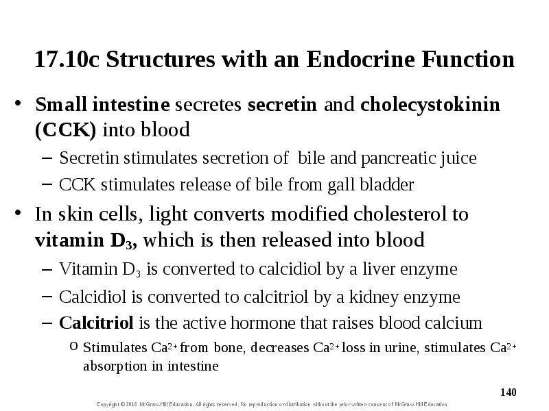
17.10c Structures with an Endocrine Function Small intestine secretes secretin and cholecystokinin (CCK) into blood Secretin stimulates secretion of...
Описание слайда:
17.10c Structures with an Endocrine Function Small intestine secretes secretin and cholecystokinin (CCK) into blood Secretin stimulates secretion of bile and pancreatic juice CCK stimulates release of bile from gall bladder In skin cells, light converts modified cholesterol to vitamin D3, which is then released into blood Vitamin D3 is converted to calcidiol by a liver enzyme Calcidiol is converted to calcitriol by a kidney enzyme Calcitriol is the active hormone that raises blood calcium Stimulates Ca2+ from bone, decreases Ca2+ loss in urine, stimulates Ca2+ absorption in intestine

Слайд 141


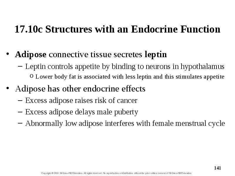
17.10c Structures with an Endocrine Function Adipose connective tissue secretes leptin Leptin controls appetite by binding to neurons in hypothalamus...
Описание слайда:
17.10c Structures with an Endocrine Function Adipose connective tissue secretes leptin Leptin controls appetite by binding to neurons in hypothalamus Lower body fat is associated with less leptin and this stimulates appetite Adipose has other endocrine effects Excess adipose raises risk of cancer Excess adipose delays male puberty Abnormally low adipose interferes with female menstrual cycle

Слайд 142


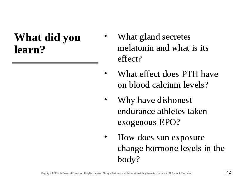
What did you learn? What gland secretes melatonin and what is its effect? What effect does PTH have on blood calcium levels? Why have dishonest...
Описание слайда:
What did you learn? What gland secretes melatonin and what is its effect? What effect does PTH have on blood calcium levels? Why have dishonest endurance athletes taken exogenous EPO? How does sun exposure change hormone levels in the body?

Слайд 143


17.11 Aging and the Endocrine System Describe how endocrine activity changes as people age.
Описание слайда:
17.11 Aging and the Endocrine System Describe how endocrine activity changes as people age.

Слайд 144


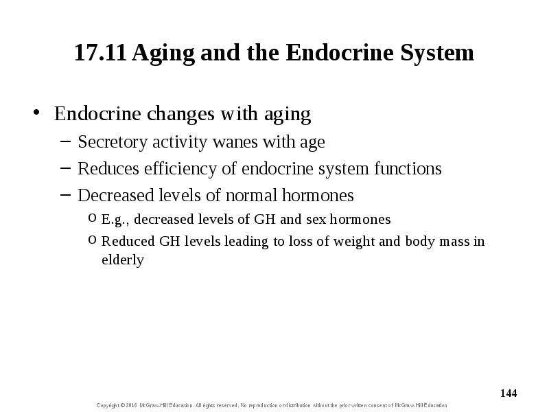
17.11 Aging and the Endocrine System Endocrine changes with aging Secretory activity wanes with age Reduces efficiency of endocrine system functions...
Описание слайда:
17.11 Aging and the Endocrine System Endocrine changes with aging Secretory activity wanes with age Reduces efficiency of endocrine system functions Decreased levels of normal hormones E.g., decreased levels of GH and sex hormones Reduced GH levels leading to loss of weight and body mass in elderly

Слайд 145


What did you learn? How does hormone replacement therapy relate to aging?
Описание слайда:
What did you learn? How does hormone replacement therapy relate to aging?


Теги chapt17 lecture2e
Похожие презентации
Mypresentation.ru
Загрузить презентацию