Описание слайда:
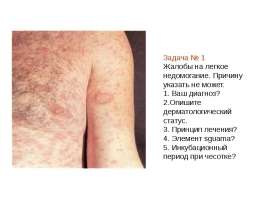
Primary syphilis After an incubation period of 2 to 6 weeks following exposure, a papule develops at the site of inoculation, which will then ulcerate into the characteristic syphilitic chancre (figure 9-11). The classic chancre is a painless, indurated ulcer with well-defined borders and a clean base. A chancre can develop on the oral (figure 11) or anorectal mucosa as well as in the genital mucosa (figure 9-10). Prior application of topical antibiotics or the use of systemic antimicrobials, may change the typical appearance of the lesion. Non-tender lymphadenopathy may be present. Primary syphilis After an incubation period of 2 to 6 weeks following exposure, a papule develops at the site of inoculation, which will then ulcerate into the characteristic syphilitic chancre (figure 9-11). The classic chancre is a painless, indurated ulcer with well-defined borders and a clean base. A chancre can develop on the oral (figure 11) or anorectal mucosa as well as in the genital mucosa (figure 9-10). Prior application of topical antibiotics or the use of systemic antimicrobials, may change the typical appearance of the lesion. Non-tender lymphadenopathy may be present. Secondary syphilis Approximately 60% to 90% of patients with untreated primary syphilis will develop manifestations of secondary syphilis. Secondary syphilis is a systemic disease that results from dissemination of the treponemes. Systemic symptoms include generalized lymphadenopathy, fever, headache, sore throat and arthralgias. Numerous clinical manifestations occur 4 to 10 weeks after the chancre disappears (or 2 to 6 months after sexual contact). These involve dermatologic (figure 12-13), central nervous system (aseptic meningitis, cranial neuropathy), ocular (iritis, uveitis or conjunctivitis), hepatic (hepatitis) and renal (immune complex glomerulonephritis) systems. The most common manifestation of secondary syphilis is the skin rash characterized by macules and papules distributed on the head and neck, the trunk and extremities including the palms and soles. The rash may be confused with pityriasis rosea, psoriasis or drug eruption. Condyloma lata are large, raised whitish lesions that are seen in warm, moist areas which occur before or soon after the rash and are highly infectious. These need to be distinguished from condyloma acuminata of human papillomavirus infections. Mucous patches are shallow, painless ulcerations that can be found on the oral or anorectal mucosa. Latent syphilis Latent syphilis is defined by reactive serology in the absence of clinical signs or symptoms. After resolution of early (primary or secondary) syphilis, mucocutaneous lesions can recur for up to 1 to 2 years in 25% of the patients. Early latent syphilis is defined as the first year from the suspected exposure when the patient is still at risk for relapse of the manifestations of secondary syphilis. Late latent syphilis is defined as a time period of one year or more after the primary infection and before the onset of tertiary syphilis. Tertiary syphilis Tertiary syphilis or late syphilis can occur after primary, secondary or latent syphilis. In the pre-antibiotic era, 25% to 40% of all patients with syphilis developed tertiary syphilis. It may present with cardiovascular manifestations, gummatous lesions or CNS disease. Cardiovascular manifestations include aortic aneurysms, aortic insufficiency or coronary stenosis. Gummatous lesions are focal inflammatory areas that can involve any organ (e.g. the liver, figure 17) but usually involve the skin (figure 15-16) and bones. Neurological disease during the tertiary stage presents as general paresis or tabes dorsalis. Neurosyphilis Infection of the CNS by the treponemes can occur at any time during the course of syphilis infection. In 15% to 40% of patients with untreated primary and secondary syphilis, T. pallidum was found in the CSF by animal inoculation studies. Treponemal invasion of the CNS during untreated early syphilis may have the following outcomes: spontaneous resolution, asymptomatic neurosyphilis (at any time during syphilis infection), acute syphilitic meningitis (in the first year), meningovascular syphilis (5 to 12 years after primary infection), and parenchymatous neurosyphilis (18 to 25 years after primary infection).